

Increased plasma neurofilament light chain concentration correlates with severity of post-mortem neurofibrillary tangle pathology and neurodegeneration
- PMID: 30626432
- PMCID: PMC6327431
- DOI: 10.1186/s40478-018-0649-3
Increased plasma neurofilament light chain concentration correlates with severity of post-mortem neurofibrillary tangle pathology and neurodegeneration
Abstract
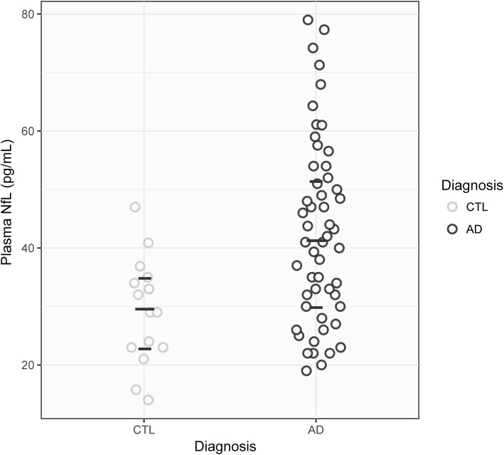
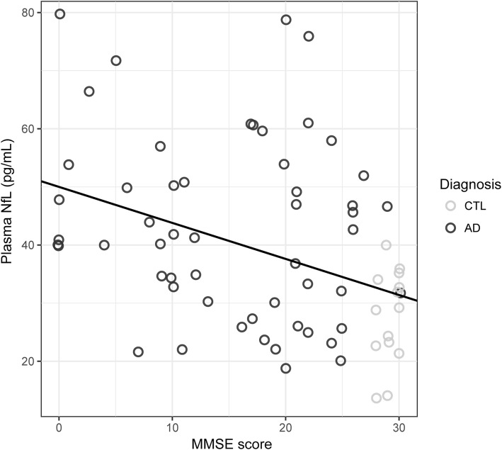
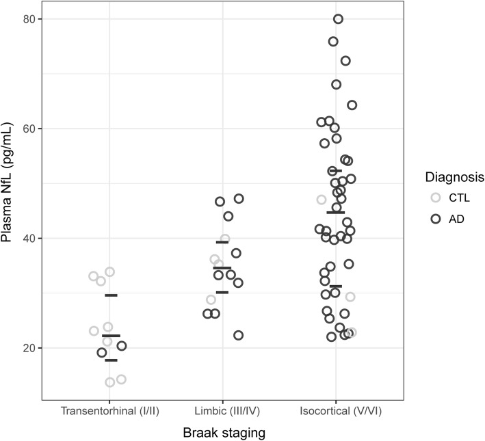
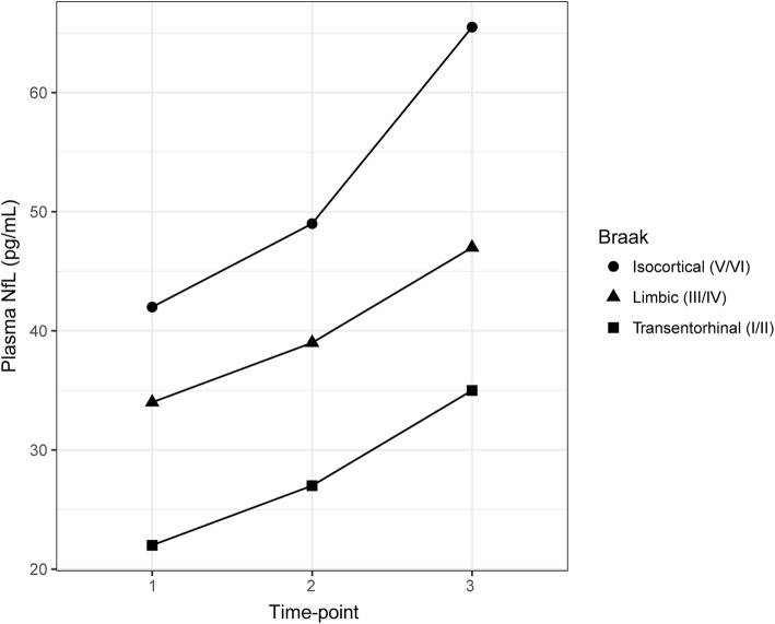
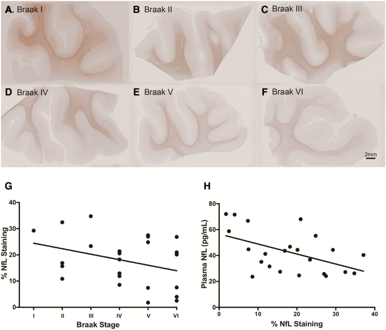
Alzheimer's disease (AD) is pathologically characterized by the accumulation of amyloid-β (Aβ) plaques, neurofibrillary tangles and widespread neuronal loss in the brain. In recent years, blood biomarkers have emerged as a realistic prospect to highlight accumulating pathology for secondary prevention trials. Neurofilament light chain (NfL), a marker of axonal degeneration, is robustly elevated in the blood of many neurological and neurodegenerative conditions, including AD. A strong relationship with cerebrospinal fluid (CSF) NfL suggests that these biomarker modalities reflect the same pathological process. Yet, the connection between blood NfL and brain tissue pathology has not been directly compared. In this study, longitudinal plasma NfL from cognitively healthy controls (n = 12) and AD participants (n = 57) were quantified by the Simoa platform. On reaching post-mortem, neuropathological assessment was performed on all participants, with additional frozen and paraffin-embedded tissue acquired from 26 participants for further biochemical (Aβ1-42, Aβ1-40, tau) and histological (NfL) evaluation. Plasma NfL concentrations were significantly increased in AD and correlated with cognitive decline, independent of age. Retrospective stratification based on Braak staging revealed that baseline plasma NfL concentrations were associated with higher neurofibrillary tangle pathology at post-mortem. Longitudinal increases in plasma NfL were observed in all Braak groupings; a significant negative association, however, was found between plasma NfL at time point 1 and both its rate of change and annual percentage increase. Immunohistochemical evaluation of NfL in the medial temporal gyrus (MTG) demonstrated an inverse relationship between Braak stages and NfL staining. Importantly, a significant negative correlation was found between the plasma NfL measurement closest to death and the level of NfL staining in the MTG at post-mortem. For the first time, we demonstrate that plasma NfL associates with the severity of neurofibrillary tangle pathology and neurodegeneration in the post-mortem brain.
Keywords: Blood biomarkers; Braak; Neurofilament light chain; Post-mortem; Tau.
Conflict of interest statement
Competing interests
KB and HZ are co-founders of Brain Biomarker Solutions in Gothenburg AB, a GU Ventures-based platform company at the University of Gothenburg. KB has served as a consultant or at advisory boards for Alzheon, Eli Lilly, Fujirebio Europe, IBL International, Novartis and Roche Diagnostics. HZ has served at advisory boards of Eli Lilly and Roche Diagnostics and has received travel support from TEVA. SL is an employee of Johnson and Johnson.
Publisher’s Note
Springer Nature remains neutral with regard to jurisdictional claims in published maps and institutional affiliations.
Figures
References
-
- Bacioglu M, Maia LF, Preische O, Schelle J, Apel A, Kaeser SA, Schweighauser M, Eninger T, Lambert M, Pilotto A, et al. Neurofilament Light Chain in Blood and CSF as Marker of Disease Progression in Mouse Models and in Neurodegenerative Diseases. Neuron. 2016;91:56–66. doi: 10.1016/j.neuron.2016.05.018. - DOI - PubMed
-
- Buerger K, Ewers M, Pirttila T, Zinkowski R, Alafuzoff I, Teipel SJ, DeBernardis J, Kerkman D, McCulloch C, Soininen H, et al. CSF phosphorylated tau protein correlates with neocortical neurofibrillary pathology in Alzheimer’s disease. Brain. 2006;129:3035–3041. doi: 10.1093/brain/awl269. - DOI - PubMed
-
- Byrne LM, Rodrigues FB, Blennow K, Durr A, Leavitt BR, Roos RAC, Scahill RI, Tabrizi SJ, Zetterberg H, Langbehn D, et al. Neurofilament light protein in blood as a potential biomarker of neurodegeneration in Huntington’s disease: a retrospective cohort analysis. Lancet Neurol. 2017;16:601–609. doi: 10.1016/S1474-4422(17)30124-2. - DOI - PMC - PubMed
-
- Chatterjee P, Goozee K, Sohrabi HR, Shen K, Shah T, Asih PR, Dave P, ManYan C, Taddei K, Chung R, et al. Association of plasma neurofilament light chain with neocortical amyloid-beta load and cognitive performance in cognitively normal elderly participants. J Alzheimers Dis. 2018;63:479–487. doi: 10.3233/JAD-180025. - DOI - PubMed
Publication types
MeSH terms
Substances
Grants and funding
LinkOut - more resources
Full Text Sources
Other Literature Sources
Medical
