. 2019 Mar;68(3):637-647.
doi: 10.2337/db18-1064. Epub 2019 Jan 9.

The Role of Prostaglandins in Disrupted Gastric Motor Activity Associated With Type 2 Diabetes

Affiliations

The Role of Prostaglandins in Disrupted Gastric Motor Activity Associated With Type 2 Diabetes

Peter J Blair et al. Diabetes. 2019 Mar.

Abstract

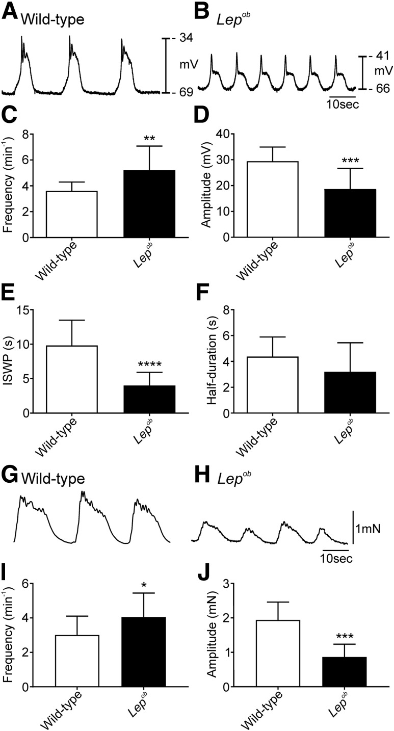
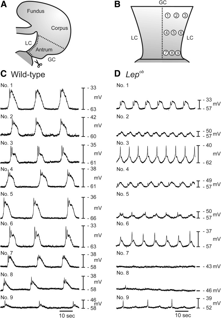
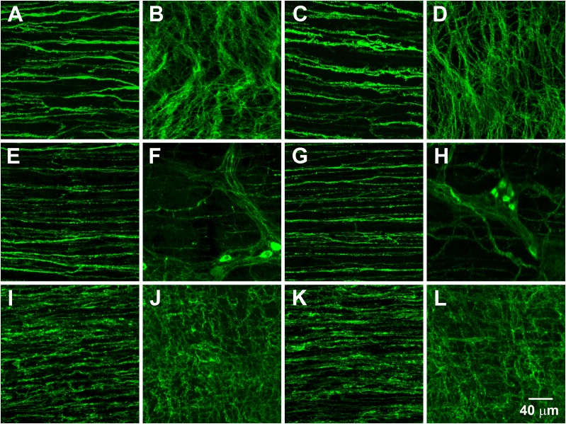
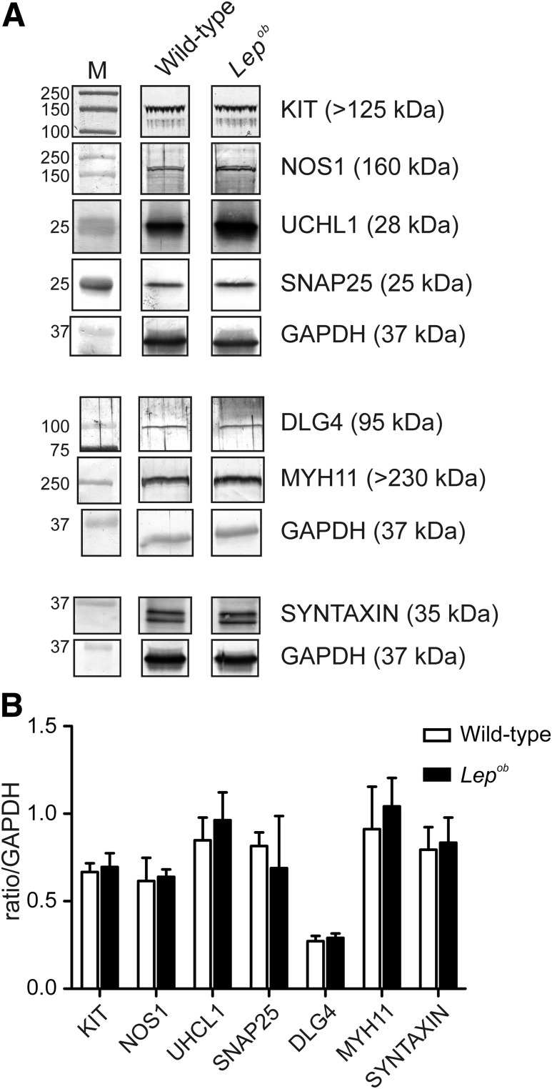
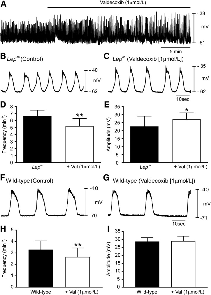
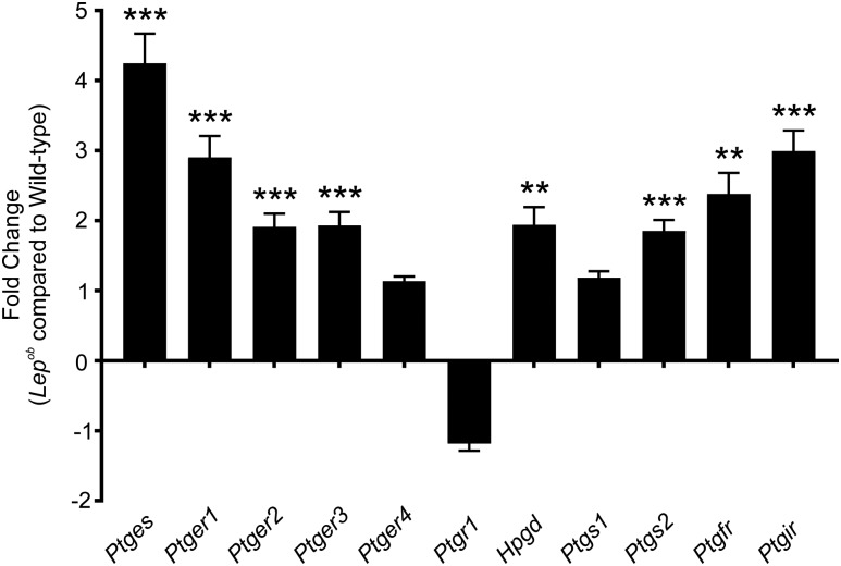
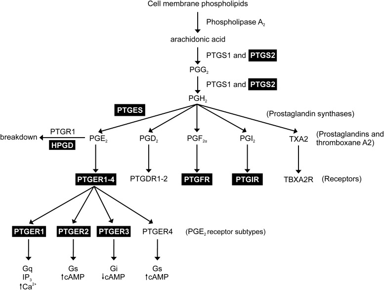
Patients with diabetes often develop gastrointestinal motor problems, including gastroparesis. Previous studies have suggested this gastric motor disorder was a consequence of an enteric neuropathy. Disruptions in interstitial cells of Cajal (ICC) have also been reported. A thorough examination of functional changes in gastric motor activity during diabetes has not yet been performed. We comprehensively examined the gastric antrums of Lepob mice using functional, morphological, and molecular techniques to determine the pathophysiological consequences in this type 2 diabetic animal model. Video analysis and isometric force measurements revealed higher frequency and less robust antral contractions in Lepob mice compared with controls. Electrical pacemaker activity was reduced in amplitude and increased in frequency. Populations of enteric neurons, ICC, and platelet-derived growth factor receptor α+ cells were unchanged. Analysis of components of the prostaglandin pathway revealed upregulation of multiple enzymes and receptors. Prostaglandin-endoperoxide synthase-2 inhibition increased slow wave amplitudes and reduced frequency of diabetic antrums. In conclusion, gastric pacemaker and contractile activity is disordered in type 2 diabetic mice, and this appears to be a consequence of excessive prostaglandin signaling. Inhibition of prostaglandin synthesis may provide a novel treatment for diabetic gastric motility disorders.


Figures

Figure 1
Figure 1
Frequency of pacemaker activity was greater, and amplitude reduced, in Lepob antrums. Representative intracellular microelectrode recordings from the antrums of wild-type (A) and Lepob (B) mice. Note the depolarized membrane potential, increased frequency, and decreased amplitude of slow waves in Lepob antrums. Time scale in B applies to both A and B. C–F: Summarized data demonstrating differences in slow wave parameters between wild-type (white bars) and Lepob antrums (black bars). Increase in slow wave frequency (C), reduction in slow wave amplitude (D), reduction in ISWP (E), and reduction in half-maximal duration of slow waves (F) (unpaired Student t test). Representative isometric force recordings from the antrums of wild-type (G) and Lepob (H) mice. Summarized data showing increased frequency (I) and reduced amplitude (J) of contractions in Lepob, as compared with wild-type (unpaired Student t test). *P ≤ 0.05; **P ≤ 0.01; ***P ≤ 0.001; ****P ≤ 0.0001.
Figure 2
Figure 2
Mapping of pacemaker activity throughout the antrums of wild-type and Lepob animals. A and B: Diagrammatic illustrations showing the gastric antrum muscle preparation and the regions from which electrical recordings were obtained. A: Antrums were separated from intact stomachs and opened along the lesser curvature (LC). B: Isolated antrums were pinned flat, the mucosa removed, and intracellular microelectrode recordings made from proximal antrum (regions 1–3), mid antrum (regions 4–6), and the terminal antrum (regions 7–9). Typical electrical activity recorded from the circular muscle layers of each of the nine regions of wild-type (C) and Lepob antrums (D). Note that membrane potentials were more depolarized and pacemaker activity was higher in frequency and decreased in amplitude in Lepob antrums compared with wild-type controls. In two regions of the Lepob antrum, no slow wave activity was recorded. GC, greater curvature.
Figure 3
Figure 3
Networks of ICC, NOS1+ nerves, and PDGFRα+ cells along the greater curvature of antrums from wild-type and Lepob animals. Representative images of ICC (A–D), NOS1+ neurons (E–H), and PDGFRα+ cells (I–L). A, E, and I show ICC, NOS1+ nerve fibers, and PDGFRα+ cells, respectively, within the circular muscle layer of a wild-type antrum. B, F, and J show ICC, NOS1+ neurons, and PDGFRα+ cells, respectively, in the myenteric plexus region of a wild-type antrum. C, G, and K show ICC, NOS1+ nerve fibers, and PDGFRα+ cells, respectively, within the circular muscle layer of a Lepob antrum. D, H, and L show ICC, NOS1+ neurons, and PDGFRα+ cells, respectively, in the myenteric plexus region of a Lepob antrum. Scale bar in L applies to all panels.
Figure 4
Figure 4
Western blot analysis for proteins relating to ICC, enteric nerves, and MYH11 cells in antrums from Lepob and wild-type animals. A: Representative Western blots comparing the expression of various proteins associated with ICC (KIT; DLG4), enteric nerves (NOS1; UCHL1, SNAP25, and syntaxin), and MYH11 in wild-type and Lepob mice, respectively. Each band is demarcated by a boundary box to illustrate that they were from different gels or different parts of the same gel. Bands are the most representative from the gastric antrums of three different animals. GAPDH was used as a control housekeeping protein to which the other proteins were compared. Three separate GAPDH blots, one from each animal, are shown. B: Western blot analysis bands from wild-type and Lepob antrums were quantified by densitometry and expressed relative to GAPDH. The summary graph demonstrates that the major proteins associated with ICC, enteric neurons, and smooth muscle were unchanged in the antrums of Lepob compared with wild-type animals (unpaired Student t test). M, marker.
Figure 5
Figure 5
PTGS2 inhibition normalizes disrupted antral pacemaker activity in Lepob antrums. A: Continuous intracellular microelectrode recording from a Lepob antrum under control conditions (no drugs) and after the addition of the PTGS2 inhibitor valdecoxib (1 μmol/L). Slow waves increased in amplitude and decreased in frequency. Intracellular recordings at a faster sweep speed before (B) and in the presence of valdecoxib (C). Traces show the changes in slow wave parameters. Summary of slow wave changes in slow wave frequency (D) and slow wave amplitude (E). Typical recordings of wild-type antrum under control conditions (F) and after the addition of valdecoxib (G). H and I: Summary of the effects of valdecoxib (Val) on slow wave frequency amplitude. Valdecoxib decreased frequency but had no effect on amplitude (paired Student t test). *P ≤ 0.05; **P ≤ 0.01.
Figure 6
Figure 6
qPCR comparing the expression of several key genes involved in prostaglandin synthesis and signaling in antrums from wild-type and Lepob mice. Bar graph depicts the fold change in prostaglandin-related gene transcripts. Several gene products were shown to be significantly upregulated in Lepob antrums compared with wild-type controls: terminal Ptges, Ptger1, Ptger2, Ptger3, Ptgs2, PGF receptor (Ptgfr), Ptgir, and Hpgd. Ptger4, Ptgr1, and Ptgs1 did not differ significantly in gene transcript expression between Lepob antrums and wild-type controls (unpaired Student t test). **P ≤ 0.01; ***P ≤ 0.001.
Figure 7
Figure 7
Diagram depicting the prostanoid synthesis pathway and the effects of PGE2 on its receptor subtypes. Highlighted boxes identify prostaglandin-related genes that are upregulated in the gastric antrums of Lepob tissues. We hypothesize that upregulation of PTGES, encoding microsomal PGE2 synthase, results in increased levels of PGE2, which in turn results in abnormal antral slow wave and contractile activity. The PTGER1–3 receptors are also upregulated and likely play a role in the altered gastric motor activity. IP3, inositol triphosphate.

References

    1. Ebert EC. Gastrointestinal complications of diabetes mellitus. Dis Mon 2005;51:620–663 - PubMed
    1. Wrzos HF, Cruz A, Polavarapu R, Shearer D, Ouyang A. Nitric oxide synthase (NOS) expression in the myenteric plexus of streptozotocin-diabetic rats. Dig Dis Sci 1997;42:2106–2110 - PubMed
    1. Stenkamp-Strahm CM, Kappmeyer AJ, Schmalz JT, Gericke M, Balemba O. High-fat diet ingestion correlates with neuropathy in the duodenum myenteric plexus of obese mice with symptoms of type 2 diabetes. Cell Tissue Res 2013;354:381–394 - PMC - PubMed
    1. Ballmann M, Conlon JM. Changes in the somatostatin, substance P and vasoactive intestinal polypeptide content of the gastrointestinal tract following streptozotocin-induced diabetes in the rat. Diabetologia 1985;28:355–358 - PubMed
    1. Ordög T, Takayama I, Cheung WK, Ward SM, Sanders KM. Remodeling of networks of interstitial cells of Cajal in a murine model of diabetic gastroparesis. Diabetes 2000;49:1731–1739 - PubMed

Publication types