. 2019 Jan 22;116(4):1299-1308.
doi: 10.1073/pnas.1816202116. Epub 2019 Jan 9.

Ribosomal protein RPL26 is the principal target of UFMylation

Affiliations

Ribosomal protein RPL26 is the principal target of UFMylation

Christopher P Walczak et al. Proc Natl Acad Sci U S A. .

Abstract

Ubiquitin fold modifier 1 (UFM1) is a small, metazoan-specific, ubiquitin-like protein modifier that is essential for embryonic development. Although loss-of-function mutations in UFM1 conjugation are linked to endoplasmic reticulum (ER) stress, neither the biological function nor the relevant cellular targets of this protein modifier are known. Here, we show that a largely uncharacterized ribosomal protein, RPL26, is the principal target of UFM1 conjugation. RPL26 UFMylation and de-UFMylation is catalyzed by enzyme complexes tethered to the cytoplasmic surface of the ER and UFMylated RPL26 is highly enriched on ER membrane-bound ribosomes and polysomes. Biochemical analysis and structural modeling establish that UFMylated RPL26 and the UFMylation machinery are in close proximity to the SEC61 translocon, suggesting that this modification plays a direct role in cotranslational protein translocation into the ER. These data suggest that UFMylation is a ribosomal modification specialized to facilitate metazoan-specific protein biogenesis at the ER.

Keywords: RPL26; UFM1; UFMylation; endoplasmic reticulum.


Conflict of interest statement

The authors declare no conflict of interest.

Figures
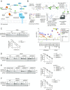
Fig. 1.
Fig. 1.
Loss of UFMylation or de-UFMylation impairs ERAD. (A) The UFMylation system shown in relation to the ER membrane bilayer. Known UFM1 conjugation or deconjugation machinery is rendered in blue and orange, respectively. Interacting partners of unknown function are shown in white with interactions denoted by dashed lines. (B) Identification of the UFMylation pathway from functional genomic analysis of substrate-selective ERAD. (Top) Forward genetic screening pipeline. Four independent genome-wide screens were iteratively conducted with the indicated GFP-tagged reporters and the data combined into a “gene effect” metric. Complete details can be found in ref. . (Bottom) Bubble plot of gene effects for UFM1-related genes and a reference set of ERAD genes. Data are from table S1 in ref. . Each of the four ERAD reporters is represented by a different colored bubble; the diameter indicates the −log P value. SYVN1 encodes HRD1, AMFR encodes GP78, and RNF139 encodes TRC8. (C) UFM1 knockout impairs turnover of CD147(CG) in K562 cells. (Top) Cells were treated with the protein synthesis inhibitor, emetine, for the indicated times before lysis. Lysates were analyzed by immunoblotting with the indicated antibodies. (Bottom) Quantification of emetine chases represented by mean ± SEM; n = 3. *P < 0.05 obtained by Student’s t test. (D) Turnover of CD147(CG) in U2OS cells is impaired by loss of UFMylation or de-UFMylation machinery. Emetine chases were performed and quantified as in C. n = 8 for wild type; n = 4 for UBA5KO, n = 5 for UFSP2KO, n = 4 for DDRGK1KO. ***P < 0.001 obtained by Student’s t test. (E) Turnover of CD147(CG) is impaired in U2OS cells expressing catalytically inactive UFSP2. Emetine chases were performed and quantified as in C; n = 3. ***P < 0.001 obtained by Student’s t test. (F) Turnover of CD147(CG) in HEK293 cells is not affected by loss of UFMylation or de-UFMylation. Emetine chases were performed and quantified as in C; n = 3. n.s., statistically not significant, P > 0.05 obtained by Student’s t test.
Fig. 2.
Fig. 2.
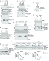
RPL26 is the principal target of UFMylation. (A) Schematic showing the genotypes of cell lines used for proteomic analysis of the UFMylome. Extent of UFMylation is represented by red intensity. (B) Proteomic analysis of the UFMylome. HEK293 cells expressing 6xHis-UFM1∆SC in UFM1KO or double knockout (DKO; UFM1KO, UBA5KO or UFM1KO, UFSP2KO) genetic backgrounds were lysed under denaturing conditions and subjected to affinity capture with Ni-NTA and LC-MS/MS analysis. TSCs for proteins identified from each affinity capture are plotted. TSCs for the nearly identical RPL26 and RPL26L1 are combined. (C) RPL26 UFMylation is undetectable in UBA5KO cells and increased in UFSP2KO cells. The indicated HEK293 cells were lysed under denaturing conditions and lysates were subjected to affinity capture with Ni-NTA. Inputs and Ni-NTA bound material were analyzed by immunoblotting with the indicated antibodies. Mobilities of mono- and di-UFMylated RPL26 are indicated. (D) RPL26 UFMylation is detected in different cell lines. Triton X-100 lysates of the indicated wild-type or UFSP2KO cell lines were analyzed by immunoblotting with the indicated antibodies. The most abundant conjugates detected with a UFM1 antibody (Bottom) comigrate with the UFMylated species of RPL26 (Top). (E) RPL26 UFMylation is enhanced in cells expressing catalytically inactive UFSP2. Triton X-100 soluble lysates of wild-type or UFSP2KO U2OS cells stably expressing the indicated UFSP2 construct were analyzed by immunoblotting with the indicated antibodies.
Fig. 3.
Fig. 3.
RPL26 is UFMylated on K132 and K134. (A) The indicated RPL26-S constructs were transfected into wild-type or UFSP2KO HEK293 cells. Cells were harvested 48 h posttransfection, lysed, and analyzed by immunoblotting with an S-tag antibody. (B) UFMylation sites on RPL26 are conserved in metazoans but absent in fungi. Sequence alignment of the C terminus of RPL26 from the indicated species was generated using Clustal Omega. RPL26 UFMylation sites identified by LC-MS/MS analysis are indicated with arrows. (C, Left) Model of the mammalian 80S ribosome bound to the SEC61/OST complex generated from Protein Data Bank (PDB) depositions 6FTG and 4UG0 (34, 36). (C, Right) View of the 60S ribosome exit tunnel opening (PDB 6FTG). RPL26 (red) is adjacent to the opening of the exit tunnel (yellow circle). K132 and K134 of RPL26 are colored blue. Dashed blue lines outline the approximate accessible surface area of UFM1 when conjugated to K134. (D) Closeup of the region encapsulated by the red box in G showing the location of K134 and K132 (blue) on the C-terminal helix of RPL26 (red). Dashed red line represents the flexible C terminus of RPL26, starting at Y135 and ending at E145, for which there is no density in the EM 3D reconstructions. (E) UFMylated RPL26 is assembled into ribosomes. Sucrose gradient sedimentation was performed on UFSP2KO HEK293 cells using a 10–50% sucrose gradient. Collected fractions were analyzed by immunoblotting (Left) and A254 absorbance (Right).
Fig. 4.
Fig. 4.
RPL26 is UFMylated and de-UFMylated at the ER membrane. (A) Knockdown of DDRGK1, UFL1, and CDK5RAP3 diminishes RPL26 UFMylation. Wild-type or UFSP2KO U2OS cells were untransfected (−), or transfected with the indicated siRNAs. Triton X-100 soluble lysates were analyzed by immunoblotting. UFM1 and RPL26 immunoblots were obtained by 4–15% and 12% SDS/PAGE, respectively. (B) DDRGK1 is required for localization of UFL1 and CDK5RAP3 to membranes. Wild-type or DDRGK1KO U2OS cells were subjected to sequential detergent extractions to isolate crude fractions before immunoblot analysis. (C) UFMylated RPL26 is enriched at membranes. As in B except wild-type cells U2OS were compared with UFSP2KO cells. (D) DDRGK1 determines the localization of UFMylated RPL26. As in C except wild-type HEK293 cells were compared with DDRGK1KO cells stably expressing C-terminally S-tagged DDRGK1 lacking its predicted transmembrane domain (DDRGK1∆TM-S). (E) Cytosolic UFMylated RPL26 assembles into ribosomes. Sucrose cushion sedimentation of lysates derived from HEK293 cells expressing only DDRGK1∆TM-S. (F) UFMylated RPL26 is detected in the membrane fraction of ODR4KO cells. The indicated HEK293 cells were fractionated with sequential detergent extractions before immunoblot analysis with the indicated antibodies. (G) UFMylated RPL26 associates with SEC61 translocons. Wild-type or UFSP2KO U2OS cells were solubilized with buffer containing 1% DMNG and subjected to immunoprecipitation with either normal rabbit IgG or antibodies raised against Sec61β. Inputs and precipitated material were divided equally and analyzed by immunoblotting. UFM1 and RPL26 immunoblots were obtained by 4–15% and 12% SDS/PAGE, respectively. (H) Loss of RPL26 UFMylation impairs ERAD of CD147(CG). (Top) Cells were treated with emetine for the indicated times before lysis and subjected to immunoblot analysis. (Bottom) Quantification represented as the mean ± SEM; n = 3. n.s., statistically not significant, P > 0.05 and *P < 0.05 obtained by Student’s t test.
Fig. 5.
Fig. 5.
Disruption of UFMylation affects transcription but not translation. (A) Scatterplots of the fold changes (knockout/wild type) in ribosome-protected reads in either cytosol or membrane fractions and of fold changes in RNA-seq reads from whole cell lysates. Pink dots represent genes that were statistically significant in the RNA-seq dataset only [false discovery rate (FDR) <0.01]. (B) Venn diagram of genes with significantly different expression (FDR <0.01) in UFM1KO and UFSP2KO HEK293 cells compared with wild type. (C) GO analysis of UFM1KO and UFSP2KO expression changes. Genes up-regulated or down-regulated in knockout cells were analyzed separately to identify statistically overrepresented GO terms (FDR <0.05, Fisher’s exact test). GO cellular component terms shared in analysis of both knockouts are displayed. The terms “extracellular matrix” and “collagen-containing ECM” were overrepresented in both up-regulated and down-regulated genes in UFSP2KO. (D) mRNA expression differences for UFSP2KO and UFM1KO cells versus wild type. Black circles indicate genes with significant differential expression in both mutants. Colored symbols highlight significantly changed genes in GO component categories related to the ECM. (E) Cumulative distribution of fold changes of genes with extracellular matrix or collagen-containing extracellular matrix annotations. P values were obtained by a Mann–Whitney–Wilcoxon U test.

References

    1. Komatsu M, et al. A novel protein-conjugating system for Ufm1, a ubiquitin-fold modifier. EMBO J. 2004;23:1977–1986. - PMC - PubMed
    1. Cai Y, Singh N, Li H. Essential role of Ufm1 conjugation in the hematopoietic system. Exp Hematol. 2016;44:442–446. - PubMed
    1. Hamilton EMC, et al. Recessive H-ABC Research Group UFM1 founder mutation in the Roma population causes recessive variant of H-ABC. Neurology. 2017;89:1821–1828. - PMC - PubMed
    1. Daniel J, Liebau E. The ufm1 cascade. Cells. 2014;3:627–638. - PMC - PubMed
    1. Tatsumi K, et al. A novel type of E3 ligase for the Ufm1 conjugation system. J Biol Chem. 2010;285:5417–5427. - PMC - PubMed

Publication types