. 2019 Mar 5;93(6):e01855-18.
doi: 10.1128/JVI.01855-18. Print 2019 Mar 15.

Natural Inhibitor of Human Cytomegalovirus in Human Seminal Plasma

Affiliations

Natural Inhibitor of Human Cytomegalovirus in Human Seminal Plasma

Sina Lippold et al. J Virol. .

Abstract

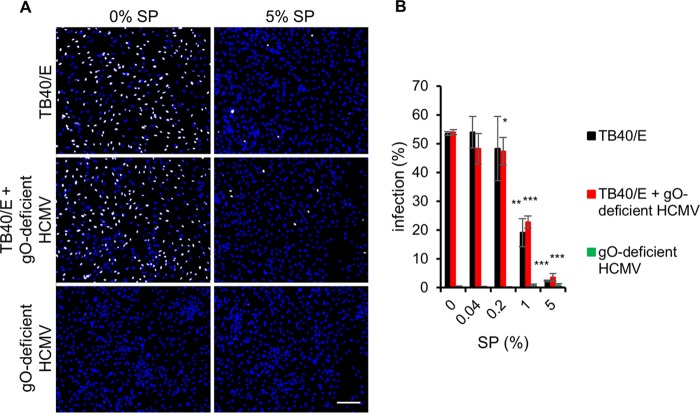
Human cytomegalovirus (HCMV) is the most frequent viral cause of congenital infections that can lead to severe birth defects. Although HCMV is frequently detected in semen and thus is potentially sexually transmitted, the role of semen in HCMV transmission is largely unclear. Here we describe that human seminal plasma (SP; the cell-free supernatant of semen) inhibits HCMV infection. The inhibition of HCMV infection was dose dependent and effective for different cell types, virus strains, and semen donors. This inhibitory effect was specific for HCMV, as herpes simplex virus 2 (HSV-2) and human immunodeficiency virus type 1 (HIV-1) infections were enhanced by SP. Mechanistically, SP inhibited infection by interfering with the attachment of virions to cells most likely via an interaction with the trimeric glycoprotein complex gH/gL/gO. Together, our findings suggest that semen contains a factor that potentially limits sexual transmission of HCMV.IMPORTANCE The role of semen in sexual transmission of human cytomegalovirus (HCMV) is currently unclear. This is surprising, as HCMV is frequently detected in this body fluid and infection is of high danger for neonates and pregnant women. In this study, we found that seminal plasma (SP) dose dependently inhibited HCMV infection. The infection inhibition was specific for HCMV, as other viruses, such as human immunodeficiency virus type 1 (HIV-1) and herpes simplex virus 2 (HSV-2), were not inhibited by SP. SP must contain a soluble, heat-resistant factor that limits attachment of HCMV particles to cells, probably by interaction with the trimeric glycoprotein complex gH/gL/gO. This novel virus-host interaction could possibly limit transmission of HCMV via semen during sexual intercourse.

Keywords: HCMV; SEVI; attachment; gH/gL/gO; glycoprotein O; human cytomegalovirus; semen; seminal amyloids; sexual transmission.


Figures

FIG 1
FIG 1
Effect of seminal plasma (SP) on HCMV, HIV-1, and HSV-2 infection. (A) HCMV (strain TB40/E, corresponding to an infection of about 20%) was incubated with the indicated concentrations of SP for 1 h at 37°C prior to infection of HFF with virus-SP mixtures for 1 h at 37°C. Infection was determined by indirect immunofluorescence staining for HCMV IE antigen 24 hpi. Blue, DAPI-positive cells; white, HCMV-positive cells. Scale bar is 200 μm. (B) Means ± SDs of relative infection from five individual infection experiments, each performed in triplicate. (C) Cytotoxicity of SP on HFF was controlled 24 h after incubation of cells with SP for 1 h at 37°C by using the MTT assay. (D and E) Means ± SDs of relative HIV-1 infection of TZM-bl cells (D) and HSV-2 infection of HFF (E) in the presence of SP from two individual experiments, each performed in triplicate. Controls (0% SP) were set to 100% and samples normalized accordingly. *, P < 0.01; **, P < 0.001; ***, P < 0.0001.
FIG 2
FIG 2
Effect of higher SP concentrations on HCMV infection. (A) Cytotoxicity of SP and heat-treated seminal plasma (hSP95°C) on HFF was investigated by the MTT assay. (B and C) HCMV (strain TB40/E, corresponding to an infection of about 20%) was incubated with SP and hSP95°C for 1 h at 37°C prior to infection of HFF with virus-SP mixtures for 1 h at 37°C. Infection was determined by indirect immunofluorescence staining for HCMV IE antigen 24 hpi. Controls (0% SP and 0% hSP95°C) were set to 100% and samples normalized accordingly. (D) Different virus doses (1×, 10×, and 100×, corresponding to infections of 20%, 50%, and 100%, respectively) of TB40/E were used in infection experiments with the indicated concentrations of hSP95°C. Given are means ± SDs from two individual experiments, each performed in triplicate. *, P < 0.01; **, P < 0.001; ***, P < 0.0001.
FIG 3
FIG 3
SP does not enhance HCMV infection at 0.1%. (A) HCMV strain TB40/E was incubated with the indicated concentrations of SP for 1 h at 37°C prior to infection of HFF with virus-SP mixtures for 24 h at 37°C. Infection was determined by indirect immunofluorescence staining for HCMV IE antigen 24 hpi. Blue, DAPI-positive cells; white, HCMV-positive cells. Scale bar is 200 μm. (B and D) HFF were infected with virus-SP mixtures for 1 h and 24 h. Infection was analyzed by indirect immunofluorescence (B) and Western blot analysis (D). (C) HFF were incubated with the indicated concentrations of SP for 1 and 24 h at 37°C. Cell viability of HFF was determined by the MTT assay. Given are means ± SDs from two individual experiments, each performed in triplicate. Controls (0% SP) were set to 100% and samples normalized accordingly. *, P < 0.01; **, P < 0.001; ***, P < 0.0001.
FIG 4
FIG 4
Effect of seminal amyloids on virus infection. (A to C) HCMV (strain TB40/E, corresponding to an infection of about 20%) was incubated with the indicated concentrations of semen-derived enhancer of virus infection (SEVI), SP, combinations of SEVI and SP, and hSP37°C (heat-treated SP devoid of seminal amyloids) for 1 h at 37°C prior to infection of HFF with mixtures for 1 h at 37°C. Infection was determined by indirect immunofluorescence staining for HCMV IE antigen 24 hpi. (A) Blue, DAPI-positive cells; white, HCMV-positive cells. Scale bar is 200 μm. (B and C) Means ± SDs from two individual experiments, each performed in triplicate. (D and E) Means ± SDs of relative infection for HIV-1 infection of TZM-bl cells (D) and for HSV-2 infection of HFF (E) in the presence of the indicated concentrations of SP or hSP37°C from two individual experiments that were performed in triplicate. Infection of the controls (0% SP) was set to 100% and samples were normalized accordingly. *, P < 0.01; **, P < 0.001; ***, P < 0.0001.
FIG 5
FIG 5
SP inhibits infection of different HCMV strains and cell types. HCMV strains TB40/E, VHL/E, AD169, Towne, and Toledo (corresponding to an infection of about 20%) were incubated with indicated concentrations of SP for 1 h at 37°C prior to infection of HFF and endothelial cells (HEC-LTT) with mixtures for 1 h at 37°C. Infection was determined by indirect immunofluorescence staining for HCMV IE antigen 24 hpi. (A) Blue, DAPI-positive cells; white, HCMV-positive cells. Scale bar is 200 μm. (B and C) Means ± SDs of relative infection from two individual experiments that were performed in triplicate. Controls (0% SP) were set to 100% and samples normalized accordingly. *, P < 0.01; **, P < 0.001; ***, P < 0.0001.
FIG 6
FIG 6
Inhibition of HCMV is a general property of human semen. HCMV (strain TB40/E, corresponding to an infection of about 50%) was incubated for 1 h at 37°C with the indicated concentrations of SP from fresh ejaculates of six donors prior to infection of HFF with mixtures for 1 h at 37°C. Infection was determined by indirect immunofluorescence staining for HCMV IE antigen 24 hpi. Given are means ± SDs of relative infection from triplicate infection. Infection of the controls (0% SP) was set to 100% and samples were normalized accordingly. *, P < 0.01; **, P < 0.001; ***, P < 0.0001.
FIG 7
FIG 7
The anti-HCMV factor in SP does not inactivate virus particles irreversibly. (A) HFF were pretreated with indicated concentrations of SP and hSP95°C (heat-treated SP) for 1 h at 37°C. Afterwards, media were removed and cells were washed and subsequently infected with TB40/E for 1 h at 37°C. (B) Cell-free supernatants of TB40/E were incubated in either medium or 5% SP and hSP95°C for 1 h at 37°C. Then viral particles were pelleted by ultracentrifugation for 2 h at 100,000 × g and 4°C. Virus pellets were resuspended in medium and used for infection experiments with HFF. (C) SP or hSP95°C in medium was processed via ultracentrifugation (100,000 × g, 2 h, and 4°C). Then supernatants were used for infection experiments with TB40/E. Cells were fixed 24 hpi and stained for HCMV IE antigen by indirect immunofluorescence. Given are means ± SDs from two individual experiments that were performed in triplicate. Infection of the controls was set to 100% and samples were normalized accordingly. *, P < 0.01; **, P < 0.001; ***, P < 0.0001.
FIG 8
FIG 8
SP inhibits attachment of HCMV virions to cell membranes. (A) Attachment of viral particles to HFF was assessed by indirect immunofluorescence staining with antibodies against the HCMV tegument protein pp150 after incubation of virus particles in the presence of SP for 1 h at 4°C. (Left) Bright field; (right) enhanced GFP (EGFP) channel (green, HCMV particles). Scale bar is 5 μm. (B) Black, quantification of attachment as in panel A with two additional conditions. Red, HFF were treated under the indicated conditions for 1 h and washed before TB40/E was added for attachment for 1 h. Green, TB40/E was added to HFF for 1 h, and cells were washed and incubated for 1 h under the indicated conditions. Given are relative numbers of attached HCMV particles per cell from 100 cells from two individual experiments. The controls (medium) were set to 100% and samples normalized accordingly. *, P < 0.01; **, P < 0.001; ***, P < 0.0001.
FIG 9
FIG 9
Infection of cells with a gO-deficient HCMV mutant is enhanced by SP. Cell-free virus particles of the gO-deficient mutant TB40-BAC4-UL74stop were incubated with the indicated concentrations of SP, hSP37°C (heat-treated SP devoid of seminal amyloids), and SEVI for 1 h at 37°C prior to infection of HEC-LTT with mixtures for 1 h at 37°C. Infection was determined by indirect immunofluorescence staining for HCMV IE antigen 24 hpi. (A) Blue, DAPI-positive cells; white, HCMV-positive cells. Scale bar is 200 μm. (B to D) Means ± SDs of relative infection from at least two individual experiments that were performed in triplicates. Infection of the controls was set to 100% and the samples were normalized accordingly. *, P < 0.01; **, P < 0.001; ***, P < 0.0001.
FIG 10
FIG 10
Ultracentrifugation of a gO-deficient HCMV does not saturate the anti-HCMV factor in SP. TB40/E, concentrated gO-deficient HCMV and a combination of both were incubated with the indicated concentrations of SP for 1 h at 37°C prior to infection of HFF with mixtures for 1 h at 37°C. Infection was determined by indirect immunofluorescence staining for HCMV IE antigen 24 hpi. (A) Blue, DAPI-positive cells; white, HCMV-positive cells. Scale bar is 200 μm. (B) Means ± SDs of infection from triplicates. *, P < 0.01; **, P < 0.001; ***, P < 0.0001.

Similar articles

Cited by

References

    1. Staras SA, Dollard SC, Radford KW, Flanders WD, Pass RF, Cannon MJ. 2006. Seroprevalence of cytomegalovirus infection in the United States, 1988–1994. Clin Infect Dis 43:1143–1151. doi:10.1086/508173. - DOI - PubMed
    1. Fowler KB, Pass RF. 2006. Risk factors for congenital cytomegalovirus infection in the offspring of young women: exposure to young children and recent onset of sexual activity. Pediatrics 118:e286–e292. doi:10.1542/peds.2005-1142. - DOI - PubMed
    1. Lanzieri TM, Kruszon-Moran D, Gambhir M, Bialek SR. 2016. Influence of parity and sexual history on cytomegalovirus seroprevalence among women aged 20–49 years in the USA. Int J Gynecol Obstet 135:82–85. doi:10.1016/j.ijgo.2016.03.032. - DOI - PMC - PubMed
    1. Rawlinson WD, Boppana SB, Fowler KB, Kimberlin DW, Lazzarotto T, Alain S, Daly K, Doutr S, Gibson L, Giles ML, Greenlee J, Hamilton ST, Harrison GJ, Hui L, Jones CA, Palasanthiran P, Schleiss MR, Shand AW, van Zuylen WJ. 2017. Congenital cytomegalovirus infection in pregnancy and the neonate: consensus recommendations for prevention, diagnosis, and therapy. Lancet Infect Dis 17:e177–e188. doi:10.1016/S1473-3099(17)30143-3. - DOI - PubMed
    1. Cannon MJ, Davis KF. 2005. Washing our hands of the congenital cytomegalovirus disease epidemic. BMC Public Health 5:70. doi:10.1186/1471-2458-5-70. - DOI - PMC - PubMed

Publication types