. 2019 Jan 10;4(1):e123431.
doi: 10.1172/jci.insight.123431.

BACE2, a conditional β-secretase, contributes to Alzheimer's disease pathogenesis

Affiliations

BACE2, a conditional β-secretase, contributes to Alzheimer's disease pathogenesis

Zhe Wang et al. JCI Insight. .

Abstract

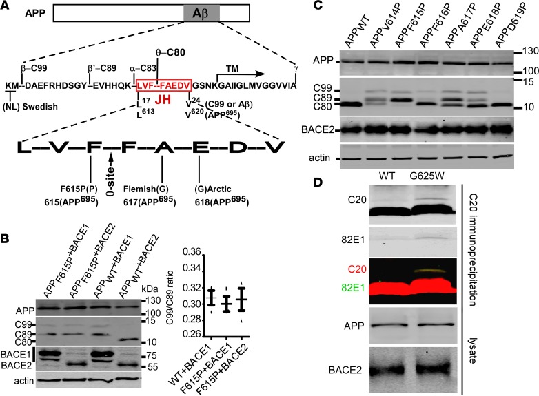
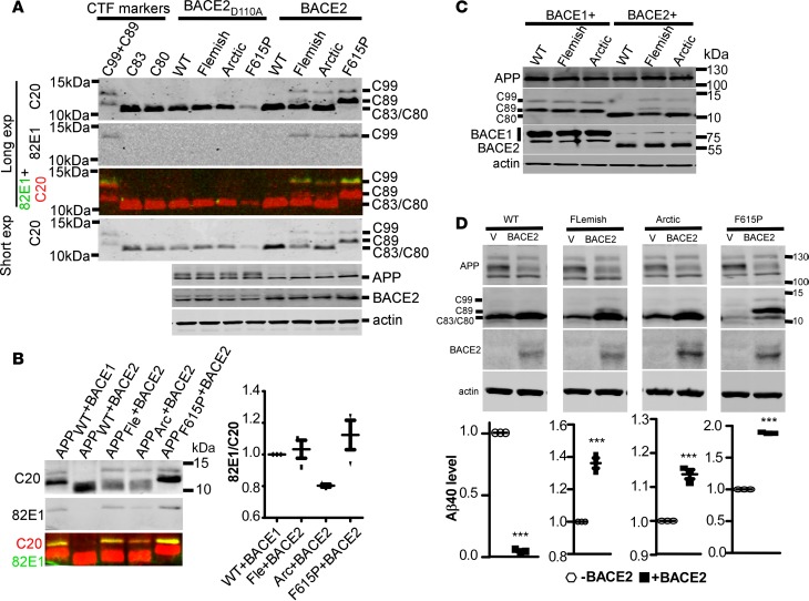
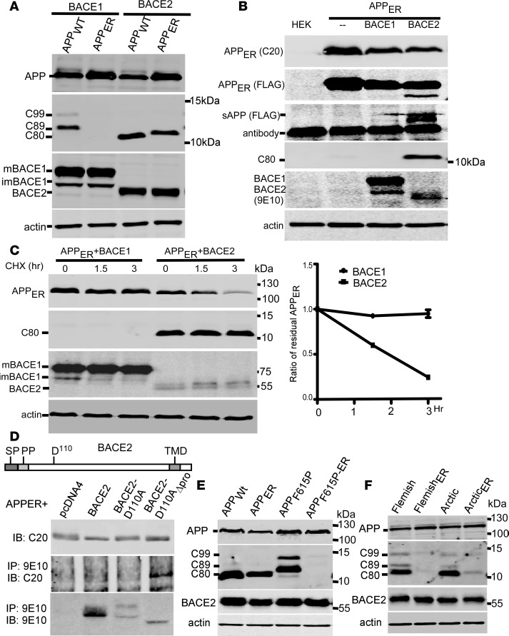
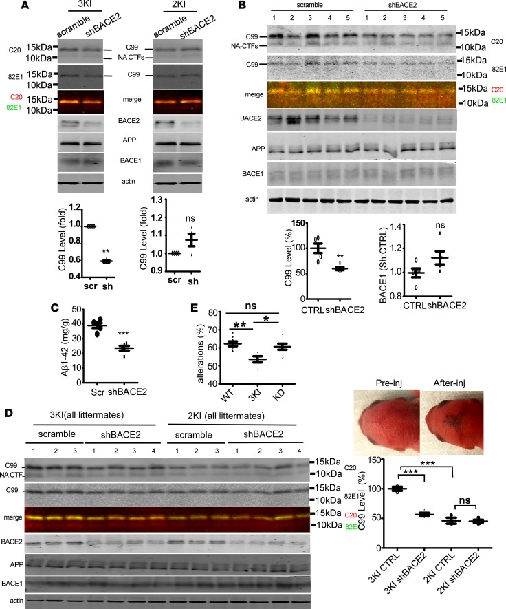
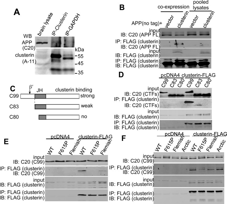
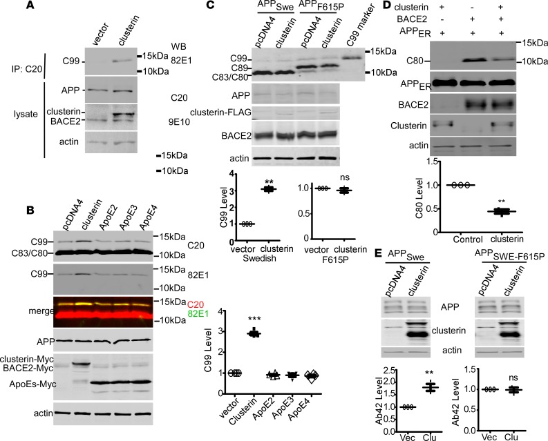
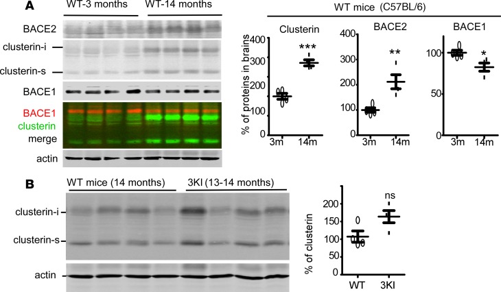
Deposition of amyloid-β protein (Aβ) to form neuritic plaques is the characteristic neuropathology of Alzheimer's disease (AD). Aβ is generated from amyloid precursor protein (APP) by β- and γ-secretase cleavages. BACE1 is the β-secretase and its inhibition induces severe side effects, whereas its homolog BACE2 normally suppresses Aβ by cleaving APP/Aβ at the θ-site (Phe20) within the Aβ domain. Here, we report that BACE2 also processes APP at the β site, and the juxtamembrane helix (JH) of APP inhibits its β-secretase activity, enabling BACE2 to cleave nascent APP and aggravate AD symptoms. JH-disrupting mutations and clusterin binding to JH triggered BACE2-mediated β-cleavage. Both BACE2 and clusterin were elevated in aged mouse brains, and enhanced β-cleavage during aging. Therefore, BACE2 contributes to AD pathogenesis as a conditional β-secretase and could be a preventive and therapeutic target for AD without the side effects of BACE1 inhibition.

Keywords: Alzheimer’s disease; Neuroscience.


Conflict of interest statement

Conflict of interest: The authors have declared that no conflict of interest exists.

Figures

Figure 1
Figure 1. Perturbation of the JH domain by proline causes BACE2-mediated β-cleavages.
(A) Schematic diagram showing the cleavage sites and mutation sites in APP. JH, juxtamembrane helix; TM, transmembrane domain. (B) APP695 wild type (APPWT) and APP F615P mutant (numbering as in APP695 form; APPF615P) were coexpressed in HEK293 cells with Myc-His–tagged BACE1 or BACE2, and cell lysates were blotted for CTFs, APP, and BACEs. Both BACE1 and BACE2 were detected using 9E10 anti-Myc antibody. Ratios of C99 to C89 were plotted. Bars represent mean ± SEM (n = 3). (C) Proline-scanning mutagenesis of the APP JH domain. All residues from V614 to D619 of APP695 were individually replaced by proline, and the mutants were coexpressed in HEK293 with BACE2. Cell lysates were immunoblotted for CTFs, APP, and BACE2. (D) APPWT and the non–membrane-binding APPG625W were coexpressed with BACE2. CTFs were enriched by IP with C20 antibody, and Western blotted with both C20 polyclonal and the C99-specific 82E1 antibody.
Figure 2
Figure 2. AD-associated APP mutations perturbed the JH domain and induced β-cleavage by BACE2.
(A) APP variants were coexpressed in HEK293 cells with BACE2 or inactive BACE2 mutant (D110A). Cell lysates were blotted for CTFs, APP, and BACE2. CTF markers expressed in HEK293 cells were loaded to indicate the mobility of CTFs. CTFs were blotted with rabbit polyclonal C20 antibody against the last 20 residues of APP, and the same membrane was also blotted with C99/Aβ(1–x)–specific mouse monoclonal antibody 82E1. The color panel is the merged images of C20 and 82E1 on the same gel. For clear visualization of C99 and C89 from APP mutants, Western blot membranes were exposed for a longer time. For the comparison of C83/C80, membranes were exposed for a shorter time. (B) APP variants were coexpressed with BACE1 or BACE2 in HEK293 cells, and CTFs were simultaneously blotted with the rabbit polyclonal C20 antibody and the mouse monoclonal 82E1 antibody. Images of C20 (red) and 82E1 (green) bands were merged in the bottom panel. Ratios of 82E1 signals to C20 signals were plotted, and comparison was by paired t tests. (C) APP wild type, Flemish, and Arctic mutants were coexpressed with either BACE1-Myc or BACE2-Myc in HEK293 cells. Cell lysates were blotted for APP, CTFs, and BACEs. (D) APPWT, Flemish, Arctic, and APPF615P mutants were coexpressed with BACE2-Myc-His in PC12 cells. Cell lysates were immunoblotted for indicated proteins and the conditioned media were measured for Aβ1–40. All bars represent mean ± SEM (n = 3). ***P < 0.001 (paired t tests). WT, APP wild type; APPFle and Fle, APP Flemish mutant; APPArc and Arc, APP Arctic mutant.
Figure 3
Figure 3. F615P, Flemish, and Arctic mutations abolished BACE2 cleavage of nascent APP.
(A) APPWT and APP with the KKXX ER retention signal (APPER) were coexpressed with BACE1 or BACE2 in HEK293 cells. CTFs and APP were blotted with C20 and BACE1 and BACE2 were probed with anti-Myc antibody. mBACE1 and imBACE1, mature and immature BACE1, respectively. (B) APPER with a FLAG tag inserted into APP after the signal peptide was coexpressed with BACE1 or BACE2 in HEK293 cells. Cell lysates were blotted for APP, CTF, and BACEs. Secreted APP (sAPP) in the conditioned media was enriched by immunoprecipitation using FLAG-agarose, and Western blotted using anti-FLAG antibody. (C) APPER was coexpressed with BACE1 or BACE2 in HEK293 cells, and treated with the translation inhibitor cycloheximide (CHX, 100 μM) for the indicated times. Full-length APPER, CTFs, and BACE2 were blotted. Immature BACE1 decreased due to ceased protein synthesis and continuous BACE1 maturation. Residual full-length APPER relative to time 0 was plotted. Curves represent mean ± SEM. (D) Schematic diagram indicates domains and the first active site in BACE2. SP, signal peptide; PP, propeptide; D110, the first active site; TMD, transmembrane domain. APPERwas coexpressed with Myc-tagged BACE2, inactive BACE2 mutant (BACE2D110A), or BACE2D110A without the propeptide (BACE2D110A-Δpro). BACE2 variants were immunoprecipitated (IP) with anti-Myc 9E10 antibody, and the precipitates were immunoblotted (IB) using the indicated antibodies. APPF615P (E) and APP Flemish and Arctic mutants (F) with the ER retention signal (APPF615P-ER, FlemishER, and ArcticER, respectively) were coexpressed with BACE2 in HEK293 cells. CTFs, APP, and BACE2 in lysates were blotted.
Figure 4
Figure 4. BACE2 suppression reduces C99 and Aβ accumulation in the brains of APP Arctic mutation–knockin mice.
(A) Primary neurons from E18 APPSwedish-Arctic-Iberian/Swedish-Arctic-Iberian–knockin mice (3KI) or APPSwedish-Iberian/Swedish-Iberian–knockin mice (2KI) were transduced with adeno-associated virus-9 (AAV9) carrying scrambled shRNA (scr) or shRNA against mouse BACE2 (sh). Cells were analyzed by Western blot for CTFs (C-terminal fragments of APP), APP, BACE2, and BACE1.NA-CTF, nonamyloidogenic C-terminal fragments of APP. (B) Viruses containing scrambled shRNA or BACE2 shRNA were intraventricularly injected into the brains of neonatal 3KI mice (all littermates, n = 5 for each). Mice were sacrificed at 11 postnatal weeks. Half brains were Western blotted for the indicated proteins. C99 on the same membrane was also blotted with C99-specific antibody 82E1, and merged with C20 blots. Nonamyloidogenic CTFs (NA-CTFs) include C89, C83, and C80. CTRL, control scrambled shRNA. (C) The other brain halves were analyzed by Aβ1–42 ELISA. ELISA readout for Aβ1–40 was no higher than background noise (n = 5). (D) AAV9 carrying scrambled shRNA or shBACE2 were intraventricularly injected into the brains of neonatal 3KI (all littermates) or 2KI (all littermates) mice. Successful injection was indicated by fast dye (blue). Mice were sacrificed 2 weeks after virus injection, and brain homogenates in RIPA buffer were Western blotted for the indicated proteins. C99 levels were expressed as percentages of C99 in scramble shRNA–injected 3KI mice. n = 4 for 3KI Ctrl, 3KI shBACE2, and 2KI shBACE2, and n = 3 for 2KI control. (E) Y-maze test for 6-month-old 3KI mice with or without BACE2 suppression. n = 8 for wild-type mice (WT), 4 for 3KI, and 8 for 3KI with suppressed BACE2 expression (KD). *P < 0.05; **P < 0.01; ***P < 0.001 by unpaired t test. ns, non-significant. All bars represent mean ± SEM.
Figure 5
Figure 5. Clusterin binds APP with intact and wild-type JH intracellularly.
(A) Coimmunoprecipitation (CoIP) of endogenous APP and clusterin from brain homogenate. Anti-GAPDH was used as control IP antibody. (B) Clusterin-FLAG and APPWT without tag were either coexpressed or separately expressed in HEK293 cells. Cells with both clusterin and APP were directly lysed (coexpression), and cells with only clusterin or APP overexpression were lysed and the lysates were combined (pooled lysates). After IP with FLAG-agarose, the precipitated proteins were blotted with C20 antibody for APP and FLAG antibody for clusterin. (C) Schematic diagram showing the position of the JH in different CTFs. (D) C99, C83, and C80 were coexpressed with clusterin-FLAG in HEK293, and the lysates were subjected to anti-FLAG CoIP. C20 antibody was used to detect CTFs in the precipitates. (E) Clusterin-FLAG was coexpressed with wild-type C99 (C99WT), F615P containing C99 (C99F615P), and Flemish mutation containing C99 (C99Fle) in HEK293 cells, and cell lysates were immunoprecipitated using anti-FLAG antibody. Precipitates were Western blotted using C20 for C99 variants and anti-FLAG for clusterin. (F) CoIP of clusterin-FLAG with APP variants in HEK293 cells. IP was by anti-FLAG antibody, and APP detection was by C20 antibody.
Figure 6
Figure 6. The binding of clusterin to the JH facilitates BACE2-mediated β-cleavages.
(A) APPWT (no tag) and BACE2 (Myc-His tagged) were expressed in HEK293 in the presence or absence (vector) of coexpressed clusterin (Myc-His tagged). CTFs were enriched by immunoprecipitation (IP) with C20 antibody, and blotted with 82E1 antibody specific for C99. (B) Clusterin or ApoEs whose binding motif in Aβ resides in the JH region as well were overexpressed in 4EB2 cells, a HEK293 cell stably expressing human APP Swedish mutant (APPSwe) and BACE2. Total CTFs were blotted with C20 antibody and C99 was also blotted with 82E1. C99s were quantified (using C20 polyclonal) and expressed as the ratio to C99 in vector-expressing cells (n = 4 replicates). ***P < 0.001 (1-way ANOVA, Tukey’s post hoc test). (C) APPSwe or APPF615P were coexpressed with either pcDNA4 or clusterin-FLAG in 4B25 cells, a HEK293 cell line stably expressing Myc-tagged human BACE2. C99 levels were expressed as the ratio to C99 in pcDNA4-APP–expressing cells (n = 3 replicates). **P < 0.01 (paired t tests). (D) Nascent APP (APPER) and BACE2 were coexpressed with pcDNA4 (vector, clusterin “–”) or clusterin (clusterin “+”) in HEK293 cells. C80 bands were quantified and plotted as the ratio to C80 in vector-expressing (clusterin “–”) cells (n = 3 replicates). **P < 0.01 (paired t tests). (E) Clusterin was coexpressed in PC12 cells with APPSwe or the APP Swedish-F615P double mutant (APPSwe-F615P). Forty-eight hours after transfection, cell lysates were blotted for the indicated proteins and conditioned media were analyzed by Aβ1–42 ELISA. The amounts of Aβ1–42 were expressed as the ratio to Aβ1–42 from cells with pcDNA4 transfection (Vec) (n = 3 replicates). **P < 0.01 (paired t tests). All bars represent mean ± SEM.
Figure 7
Figure 7. Altered expression of clusterin, BACE1, and BACE2 in aged brains.
(A) Brains from C57BL/6 mice at the age of 3 months and 14 months were homogenized in RIPA buffer and the brain lysates were Western blotted for clusterin, BACE1, and BACE2. Protein bands were quantified and plotted. For the quantification of clusterin, the intracellular form (clusterin-i) and the secreted form (clusterin-s) were combined. (B) Brain lysates of C57BL/6 mice at the age of 14 months and 13- to 14-month-old 3KI (APPSwedish-Arctic-Iberian/Swedish-Arctic-Iberian) were blotted for clusterin. Both forms of clusterin were quantified and combined for plotting. *P < 0.05; **P < 0.01; ***P < 0.001 (paired t tests). ns, non-significant. All bars represent mean ± SEM.

References

    1. Kang Y, et al. Nutritional deficiency in early life facilitates aging-associated cognitive decline. Curr Alzheimer Res. 2017;14(8):841–849. - PubMed
    1. Zhang Y, Song W. Islet amyloid polypeptide: Another key molecule in Alzheimer’s pathogenesis? Prog Neurobiol. 2017;153:100–120. doi: 10.1016/j.pneurobio.2017.03.001. - DOI - PubMed
    1. Zhang S, et al. BACE1 cleavage site selection critical for amyloidogenesis and Alzheimer’s pathogenesis. J Neurosci. 2017;37(29):6915–6925. doi: 10.1523/JNEUROSCI.0340-17.2017. - DOI - PMC - PubMed
    1. Zhang S, et al. A presenilin-1 mutation causes Alzheimer disease without affecting Notch signaling [published online ahead of print]. Mol Psychiatry. doi: 10.1038/s41380-018-0101-x. - PubMed
    1. Zeng J, et al. Marginal vitamin A deficiency facilitates Alzheimer’s pathogenesis. Acta Neuropathol. 2017;133(6):967–982. doi: 10.1007/s00401-017-1669-y. - DOI - PubMed