Review
. 2019 May 15;58(10):1433-1441.
doi: 10.2169/internalmedicine.1948-18. Epub 2019 Jan 10.

Sclerosing Angiomatoid Nodular Transformation of the Spleen: Lessons from a Rare Case and Review of the Literature

Affiliations
Review

Sclerosing Angiomatoid Nodular Transformation of the Spleen: Lessons from a Rare Case and Review of the Literature

Ryohei Nomura et al. Intern Med. .

Abstract

Sclerosing angiomatoid nodular transformation (SANT) of the spleen is an extremely rare benign lesion. We herein report a case of asymptomatic SANT of the spleen in a middle-aged woman with early breast carcinoma and an undiagnosed splenic mass, which was successfully treated by laparoscopic splenectomy and diagnosed postoperatively. We also review the literature on SANT to help make knowledge more accessible when clinicians encounter a splenic tumor. The present case taught us the following lesson: the presence of a splenic lesion during follow-up for malignancy is not always indicative of metastasis. Therefore, SANT should be considered in the differential diagnosis.

Keywords: case report; laparoscopic splenectomy; malignancy; sclerosing angiomatoid nodular transformation; spleen.


Conflict of interest statement

The authors state that they have no Conflict of Interest (COI).

Figures

Figure 1.
Figure 1.
Abdominal CT revealed a solid lesion (white arrowheads) measuring 40 mm in diameter in the periphery of the spleen (A). The lesion was less contrast-enhanced than the spleeparenchyma in the early and portal phases (B). The densities of the mass and the surrounding spleen were similar in the delayed phase (C).
Figure 2.
Figure 2.
Consistent with CT imaging, the lesion (white arrowheads) showed almost the same intensity as the splenic parenchyma on both T1- and T2-weighted MRI (A, B) and was slowly but progressively filled centripetally with contrast medium 10 minutes after injection (C). The mass reached a similar intensity to the background spleen (D).
Figure 3.
Figure 3.
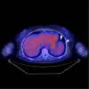
PET-CT showed no significant avidity for FDG in the splenic mass (white arrowheads).
Figure 4.
Figure 4.
The resected specimen of the spleen is shown. The well-circumscribed solitary lesion, measuring 40 mm in diameter, contained a central white stellate structure, which is characteristic of SANT.
Figure 5.
Figure 5.
Hematoxylin and Eosin staining revealed multiple angiomatoid nodules separated by fibrous or fibrosclerotic stroma (A). Proliferation of both collagen fiber (B) and microvessels was observed, with inflammatory cell infiltration (C).
Figure 6.
Figure 6.
On immunohistochemical staining, three types of vessels were observed, which is characteristic of SANT: (1) sinusoid type, both CD8- and CD31-positive but CD34-negative vessels (A); (2) small vein type, CD31-positive but CD8- and CD34-negative vessels (B); and (3) cord capillary type, both CD34- and CD31-positive but CD8-negative vessels (C). In the stroma, myofibroblastic spindle cells had proliferated, showing positivity for α-smooth muscle actin (D). These immunohistochemical findings were definitive in confirming the exact diagnosis.

References

    1. Krishnan J, Frizzera G. Two splenic lesions in need of clarification: hamartoma and inflammatory pseudotumor. Semin Diagn Pathol 20: 94-104, 2003. - PubMed
    1. Martel M, Cheuk W, Lombardi L, Lifschitz-Mercer B, Chan JK, Rosai J. Sclerosing angiomatoid nodular transformation (SANT): report of 25 cases of a distinctive benign splenic lesion. Am J Surg Pathol 28: 1268-1279, 2004. - PubMed
    1. Thipphavong S, Duigenan S, Schindera ST, Gee MS, Philips S. Nonneoplastic, benign, and malignant splenic diseases: cross-sectional imaging findings and rare disease entities. AJR Am J Roentgenol 203: 315-322, 2014. - PubMed
    1. Wang TB, Hu BG, Liu DW, Gao ZH, Shi HP, Dong WG. Sclerosing angiomatoid nodular transformation of the spleen: a case report and literature review. Oncol Lett 12: 928-932, 2016. - PMC - PubMed
    1. Union for International Cancer Control In: TNM Classification of Malignant Tumors. 8th ed Brierley JD, Gospodarowicz MK, Wittekind Ch, Eds. Wiley & Blackwell, Hoboken, USA, 2017: 151-158.