Review
. 2019 Jan;20(1):50-57.
doi: 10.3348/kjr.2018.0325. Epub 2018 Dec 27.

Four-Dimensional Thoracic CT in Free-Breathing Children

Affiliations
Review

Four-Dimensional Thoracic CT in Free-Breathing Children

Hyun Woo Goo. Korean J Radiol. 2019 Jan.

Abstract

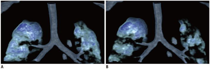
In pediatric thoracic CT, respiratory motion is generally treated as a motion artifact degrading the image quality. Conversely, respiratory motion in the thorax can be used to answer important clinical questions, that cannot be assessed adequately via conventional static thoracic CT, by utilizing four-dimensional (4D) CT. However, clinical experiences of 4D thoracic CT are quite limited. In order to use 4D thoracic CT properly, imagers should understand imaging techniques, radiation dose optimization methods, and normal as well as typical abnormal imaging appearances. In this article, the imaging techniques of pediatric thoracic 4D CT are reviewed with an emphasis on radiation dose. In addition, several clinical applications of pediatric 4D thoracic CT are addressed in various thoracic functional abnormalities, including upper airway obstruction, tracheobronchomalacia, pulmonary air trapping, abnormal diaphragmatic motion, and tumor invasion. One may further explore the clinical usefulness of 4D thoracic CT in free-breathing children, which can enrich one's clinical practice.

Keywords: 4D CT; Air trapping; Airway; CT imaging techniques; Chest CT; Child; Diaphragm motion; Lung densitometry; Tracheobronchomalacia; Tumor invasion.


Conflict of interest statement

Conflicts of Interest: The author has no financial conflicts of interest.

Figures

Fig. 1
Fig. 1. Comparison between intermittent and continuous scan modes of 4D pediatric thoracic CT.
Six intermittent and nine continuous sequential scans are performed in 2 seconds. At same 80 kV and 10 mAs, DLP value of continuous scan mode is 16 mGy·cm, which is four-times higher than that of intermittent mode (4 mGy·cm). DLP = dose length product, 4D = four-dimensional
Fig. 2
Fig. 2. Normal upper airway in 1-month-old boy with vascular ring.
Sagittal inspiratory (A) and expiratory (B) volume-rendered CT images demonstrate normal appearance of upper airway. U separating nasopharynx from oropharynx is noted. U contributes to velopharyngeal closure with other velopharyngeal muscles that is indispensable for normal swallowing and speaking. As shown in these merged volume-rendered images, bone structures can be added to airway structures to facilitate anatomical correlations. To note, upper airway dimension diminishes normally from inspiration to expiration. Patient was sucking on pacifier (asterisks) during CT examination. U = uvula
Fig. 3
Fig. 3. Dynamic pharyngeal obstruction in 2-month-old girl with complete atrioventricular septal defect.
Sagittal inspiratory (A) and expiratory (B) volume-rendered CT images reveal severe dynamic expiratory narrowing (arrow) of pharynx. E = epiglottis, L = larynx, T = tongue
Fig. 4
Fig. 4. Fixed obstruction of entire pharynx in 9-month-old boy with repaired hypoplastic left heart syndrome.
Oblique coronal inspiratory (A) and expiratory (B) volume-rendered CT images show diffuse, fixed pharyngeal narrowing (arrows). C. Axial contrast-enhanced CT image reveals soft tissue thickening causing pharyngeal narrowing (arrows).
Fig. 5
Fig. 5. Glossoptosis in 1-month-old boy with repaired diaphragmatic hernia and multiple congenital anomalies.
A. Neck lateral radiograph shows abnormal posterior displacement of tongue causing pharyngeal obstruction (arrows). Sagittal thick-slab inspiratory (B) and expiratory (C) CT images demonstrate that pharynx is patent during inspiration but completely obliterated (arrows) during expiration. A = anterior, P = posterior
Fig. 6
Fig. 6. Normal larynx in 3-month-old boy with tetralogy of Fallot and suspected laryngomalacia.
Axial inspiratory (A) and expiratory (B) thick-slab CT images show normal laryngeal appearance during respiration.
Fig. 7
Fig. 7. Laryngomalacia in 1-month-old girl with tetralogy of Fallot and inspiratory stridor.
A. Axial inspiratory thick-slab minimum intensity projection CT image shows collapsed larynx due to severe laryngomalacia. B. Axial thick-slab minimum intensity projection CT image obtained between inspiration and expiration illustrates typical omega-shaped epiglottis (arrows) caused by inward collapse of aryepiglottic folds. C. Axial expiratory thick-slab minimum intensity projection CT image displays intact laryngeal shape. D. Sagittal inspiratory volume-rendered CT image illustrates excessive inspiratory collapse of supraglottic airway (arrows) due to severe laryngomalacia. E. Sagittal inspiratory volume-rendered CT image shows improvement of laryngomalacia after epiglottopexy.
Fig. 8
Fig. 8. Tracheomalacia in 2-month-old boy with tetralogy of Fallot after esophagoesophagostomy for esophageal atresia and ligated tracheoesophageal fistula.
Coronal inspiratory (A) and expiratory (B) volume-rendered CT images show severe expiratory tracheal collapse (long arrows) indicating tracheomalacia at thoracic inlet level. Longitudinal extent of focal tracheomalacia is nicely illustrated on expiratory-phase 4D CT image (B). Remnant of ligated tracheoesophageal fistula (short arrows) is noted.
Fig. 9
Fig. 9. Pulmonary air trapping in 14-day-old boy with repaired complete transposition of great arteries and vascular ring.
Coronal inspiratory (A) and expiratory (B) volume-rendered CT images reveal multiple lung lesions with air trapping, suggesting small airway disease. Characteristic mosaic lung attenuation is shown on expiratory-phase 4D CT image (B).

References

    1. Lee EY, Mason KP, Zurakowski D, Waltz DA, Ralph A, Riaz F, et al. MDCT assessment of tracheomalacia in symptomatic infants with mediastinal aortic vascular anomalies: preliminary technical experience. Pediatr Radiol. 2008;38:82–88. - PubMed
    1. Long FR, Castile RG, Brody AS, Hogan MJ, Flucke RL, Filbrun DA, et al. Lungs in infants and young children: improved thin-section CT with a noninvasive controlled-ventilation technique--initial experience. Radiology. 1999;212:588–593. - PubMed
    1. Goo HW, Allmendinger T. Combined electrocardiography- and respiratory-triggered CT of the lung to reduce respiratory misregistration artifacts between imaging slabs in free-breathing children: initial experience. Korean J Radiol. 2017;18:860–866. - PMC - PubMed
    1. Goo HW. Combined prospectively electrocardiography- and respiratory-triggered sequential cardiac computed tomography in free-breathing children: success rate and image quality. Pediatr Radiol. 2018;48:923–931. - PubMed
    1. Lin L, Bivard A, Krishnamurthy V, Levi CR, Parsons MW. Whole-brain CT perfusion to quantify acute ischemic penumbra and core. Radiology. 2016;279:876–887. - PubMed

LinkOut - more resources