Review
. 2018 Dec 21:12:106.
doi: 10.3389/fncir.2018.00106. eCollection 2018.

Neuropeptide and Small Transmitter Coexistence: Fundamental Studies and Relevance to Mental Illness

Affiliations
Review

Neuropeptide and Small Transmitter Coexistence: Fundamental Studies and Relevance to Mental Illness

Tomas Hökfelt et al. Front Neural Circuits. .

Abstract

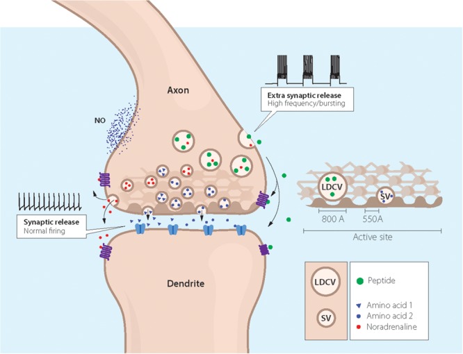
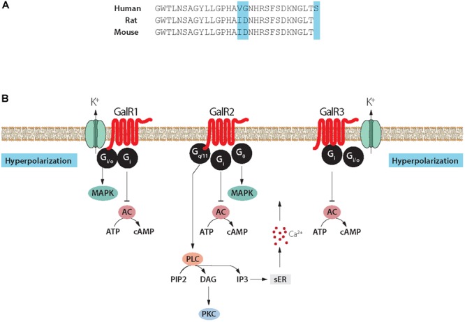
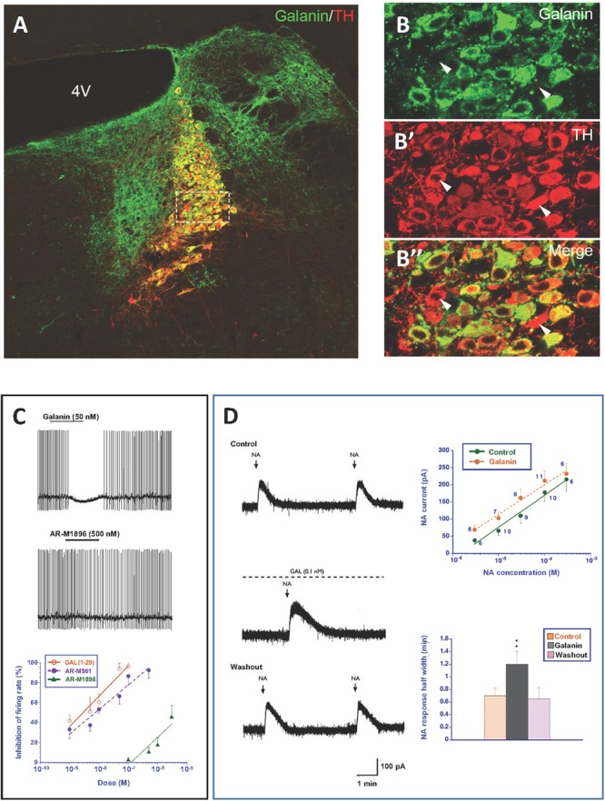
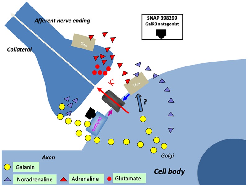
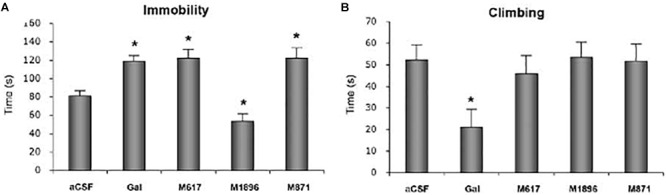
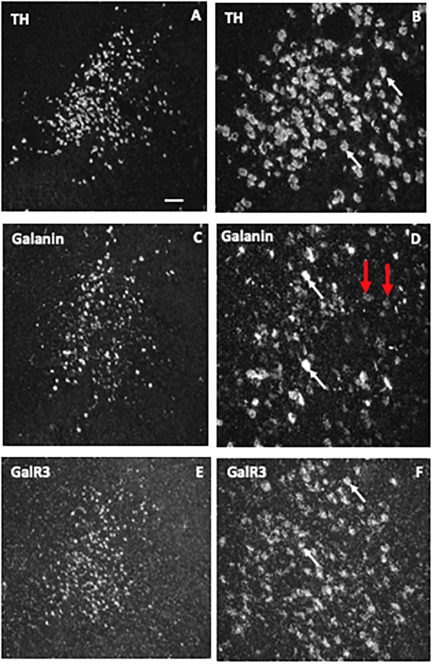
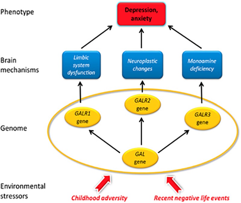
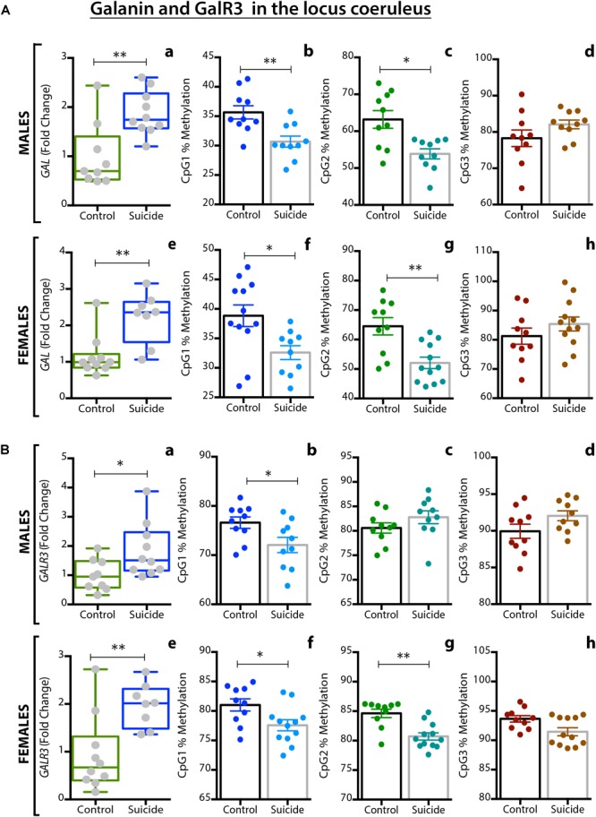
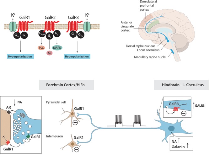
Neuropeptides are auxiliary messenger molecules that always co-exist in nerve cells with one or more small molecule (classic) neurotransmitters. Neuropeptides act both as transmitters and trophic factors, and play a role particularly when the nervous system is challenged, as by injury, pain or stress. Here neuropeptides and coexistence in mammals are reviewed, but with special focus on the 29/30 amino acid galanin and its three receptors GalR1, -R2 and -R3. In particular, galanin's role as a co-transmitter in both rodent and human noradrenergic locus coeruleus (LC) neurons is addressed. Extensive experimental animal data strongly suggest a role for the galanin system in depression-like behavior. The translational potential of these results was tested by studying the galanin system in postmortem human brains, first in normal brains, and then in a comparison of five regions of brains obtained from depressed people who committed suicide, and from matched controls. The distribution of galanin and the four galanin system transcripts in the normal human brain was determined, and selective and parallel changes in levels of transcripts and DNA methylation for galanin and its three receptors were assessed in depressed patients who committed suicide: upregulation of transcripts, e.g., for galanin and GalR3 in LC, paralleled by a decrease in DNA methylation, suggesting involvement of epigenetic mechanisms. It is hypothesized that, when exposed to severe stress, the noradrenergic LC neurons fire in bursts and release galanin from their soma/dendrites. Galanin then acts on somato-dendritic, inhibitory galanin autoreceptors, opening potassium channels and inhibiting firing. The purpose of these autoreceptors is to act as a 'brake' to prevent overexcitation, a brake that is also part of resilience to stress that protects against depression. Depression then arises when the inhibition is too strong and long lasting - a maladaption, allostatic load, leading to depletion of NA levels in the forebrain. It is suggested that disinhibition by a galanin antagonist may have antidepressant activity by restoring forebrain NA levels. A role of galanin in depression is also supported by a recent candidate gene study, showing that variants in genes for galanin and its three receptors confer increased risk of depression and anxiety in people who experienced childhood adversity or recent negative life events. In summary, galanin, a neuropeptide coexisting in LC neurons, may participate in the mechanism underlying resilience against a serious and common disorder, MDD. Existing and further results may lead to an increased understanding of how this illness develops, which in turn could provide a basis for its treatment.

Keywords: allostatic load; epigenetics; galanin; locus coeruleus; major depression disorder; neuropeptides; resilience.


Figures

FIGURE 1
FIGURE 1
Immunofluorescence micrographs of the guinea-pig inferior mesenteric ganglion (A,B) and electron micrographs from different types of nerve endings (C–E). (A,B) Two adjacent sections incubated with antibodies to somatostatin (A) and the noradrenaline (NA) synthesizing enzyme dopamine ß-hydroxylase (DBH) (B). The majority of the principal ganglion cells are somatostatin-positive, whereas the small intensely fluorescent (SIF) cells (asterisk) lack the peptide. Virtually all ganglion cells and the SIF cells are DBH-positive, i.e., are noradrenergic. (C–E) Examples of transmitter storage in nerve endings based on or immunohistochemistry (C,E) or potassium permanganate fixation (D). (D) In sympathetic nerve endings NA (black precipitate) is stored in both (small) synaptic vesicles and large dense core vesicle (LDCVs) (arrow). Note that content varies between vesicles, both in the synaptic and LDCVs. (C) Substance P, a neuropeptide (black precipitate), in a sensory nerve ending in the monkey dorsal horn, is stored exclusively in LDCVs, all synaptic vesicles are empty. (E) Peptide and glutamate co-storage and coexistence in the dorsal horn of the rat spinal cord based on immunogold immunohistochemistry. Substance P/CGRP is detected with 10/20 nm gold particles and glutamate with 5 nm gold particles. Note that substance P and CGRP can be stored within the same LDCV (left box, magnified in E’). Staining for glutamate is restricted to synaptic vesicles (right box, magnified in E”). The results suggest that glutamate, a small molecule transmitter, is not stored in LDCVs in sensory nerve endings, and release of peptide and amino acid may be separate events. This contrasts NA (see D). Bars: 40 μm, for (A,B); 100 nm for (C,D); 250 nm for (E). (A,B) From Hokfelt et al. (1977a). (C) From DiFiglia et al. (1982), with permission. (D,E) Courtesy of Dr. A. Merighi (cf., Merighi, 2002).
FIGURE 2
FIGURE 2
Coexistence and subcellular distribution of neuropeptide Y (NPY) and noradrenaline (NA) in the rat vas deferens. (A,B) Immunohistochemical visualization of NPY- (A) and tyrosine hydroxylase (TH)-(B) positive nerve terminals in adjacent sections. Overlapping, dense NPY and noradrenergic networks are seen in the muscle layer. Note sparse NPY-only positive nerves (arrow) in the subepithelial region, possibly cholinergic nerves. (C) Electron microscopic micrograph of several nerve terminal profiles in the muscle layer after potassium permanganate (KMnO4) fixation, showing small synaptic vesicles with a dense core and LDCVs. The dense core indicates presence of NA both in the synaptic and LDCVs (cf. Figure 1D). No profiles without small vesicle with a dense core are seen, suggesting a pure adrenergic innervation of the muscle layer. (D,E) Subcellular distribution of NA (x) and NPY (o) in a density gradient of rat vas deferens. There is only one peak for NPY (fraction 7; E), whereas there are two peaks for NA (fraction 5 and 7), tentatively representing synaptic vesicles and LDCVs, respectively. Note many LDCVs (arrows), as well as many vesicles of the same size but without dense core (double-headed arrow). The peptide is only present in the heavy fraction (in agreement with Figures 1C,E), whereas NA is present also in the light one (in agreement with Figure 1D). On the abscissa, totally recovered sedimentable substance is given as picomoles per milliliter after centrifugation at 145,000 × gmax for 45 min. On the ordinate, density gradient fractions 1–10 are given, corresponding to the following sucrose molarities: 1 (0.26 M), 2 (0.32 M), 3 (0.47 M), 4 (0.56 M), 5 (0.69 M), 6 (0.74 M), 7 (0.84 M), 8 (0.91 M), 9 (0.98 M), 10 (1.2 M). Recoveries of NA = 70%, of NPY = 65%, and of protein = 87%. Reprinted from Fried et al. (1985), with permission.
FIGURE 3
FIGURE 3
Cartoon showing coexistence of a neuropeptide with classic and ‘unconventional’ neurotransmitters in a nerve ending synapsing on a dendrite. Two types of storage vesicles are shown: synaptic vesicles (diameter 500 Å) storing classic transmitters (e.g., 5-HT, NA, GABA or glutamate), mainly released at synapses; large dense core vesicles (LDCVs) storing neuropeptides and, in amine neurons NA or 5-HT. The peptides are in general released extrasynaptically (“volume transmission”), when neurons fire with high frequency or in bursts. Peptide receptors are essentially extrasynaptic or presynaptic, whereas ligand-gated receptors are mostly localized in the postsynaptic membrane. ‘Gaseous’ (e.g., nitric oxide, NO) and other non-conventional transmitters are not stored in vesicles, but are generated upon demand (Snyder and Ferris, 2000). The presynaptic grid, an egg basket-like structure, originally described by Pfenninger et al. (1969), is indicated in the nerve ending and high-lighted to the right. Note that the LDCV does not fit into the grid and thus cannot attach to the presynaptic membrane for release. In contrast, there is room for the synaptic vesicle. This supports the concept that peptides are mostly not released into the synaptic cleft. Drawing by Mattias Karlen. Modified from Pfenninger et al. (1969),Lundberg and Hokfelt (1983), and Lang et al. (2015).
FIGURE 4
FIGURE 4
(A) Structure of galanin in three species. Galanin is composed of 29 amino acids in most species, except humans (30 amino acids). Note conservation of N-terminal portion. (B) Signaling pathways of galanin receptor subtypes. Galanin, via GalR1 and GalR3, opens potassium channels leading to membrane hyperpolarization. Galanin can via GalR2 activate PLC resulting in generation of IP3, release of Ca2+ from the smooth endoplasmic reticulum, opening of Ca2+ channels and eventually transmitter release. AC, adenylate cyclase; cAMP, 3′, 5′-cyclic adenosine monophosphate; DAG, diacylglycerol; K+, G-protein-regulated inwardly rectifying potassium channel; sER, smooth endoplasmic reticulum; IP3, inositol triphosphate; PIP2, phosphatidylinositol bisphosphate; PKC, protein kinase C; PLC, phospholipase C. Modified from Iismaa and Shine (1999) and Lang et al. (2015). Drawing by Mattias Karlén.
FIGURE 5
FIGURE 5
(A–B”) Immunofluorescence micrographs of the dorsal pontine periventricular region of mouse after double-staining of a section with antibodies to galanin (green) and tyrosine hydroxylase (TH) (red), the rate-limiting enzyme for catecholamine synthesis and thus a marker for NA neurons. Note that both antibodies stain neurons in the locus coerulus (LC) (B,B’), whereby many (yellow, B”), but not all TH-positive neurons express galanin [arrowheads point to TH-only neurons (red), apparently lacking galanin] (B’). Galanin is also present in many structures outside the LC. Colchicine treated animal. Courtesy Joanne Bakker and Mingdong Zhang. Bar for (A) 200 μm, for (B–B”) 20 μm. (C) Effect of galanin and the GalR2 agonist AR-M1896 on LC neurons (upper two traces), and the dose–response curves of galanin (red), the AR-M1896 (green) and the mixed GalR1-GalR2 M961 agonist (magenta) (lower trace). Note strong hyperpolarization of galanin and a less strong effect of M961, whereas that AR-M1896 hardly causes any effect at all. From Ma et al. (2001). (D, left panel) Effect of galanin on the response of LC neurons to NA. NA (applied from a pipette at the arrowhead) induces a persistent outward current (upper trace). When galanin (0.1 nM) is present, the NA-induced outward current is enhanced, and the duration is prolonged (middle trace). After wash out of galanin, the amplitude and duration of the NA response was similar to that seen before galanin administration (lower trace). (D, right panel) Effect of galanin on dose-response (upper figure) and duration (lower figure) of NA. The NA dose-response curve is shifted to the left, when galanin (0.1 nM) is present (upper figure). The duration of the NA-induced current is increased in the presence of galanin (lower figure). ∗∗P < 0.01. From Xu et al. (2001) with permission.
FIGURE 6
FIGURE 6
Cartoon showing several transmitters and signaling pathways in the locus coeruleus (LC) (part of a cell body with initial axon and an afferent nerve ending and a possible axon collateral). A noradrenergic LC neuron co-expresses galanin (yellow LDCVs) originating in the Golgi complex. The peptide in the LDCVs is, after transport to the somatic and dendritic cell membrane, released by exocytosis. Galanin then acts on inhibitory autoreceptors (GalR1/R3), opening potassium channels, in this way attenuating noradrenaline (NA) release in the forebrain. Galanin at low concentrations enhance the alpha2A mediated inhibition of the LC neuron (by an unknown mechanism). Galanin could also be released from collaterals. The GalR3 antagonist (SNAP 398299) may exert an antidepressive action by disinhibiting the LC neuron and restituting forebrain NA levels. With regard to small transmitters, NA (purple triangles) can be released from soma-dendrites and collaterals, acting on somato-dendritic, postsynaptic and presynaptic alpha2A receptors. The afferent nerve ending originates from C1 neurons which are glutamatergic (red dots) and co-release adrenaline (red triangles). Also adrenaline can act on the alpha2A receptors. The basis for this cartoon is animal experiments, and in the case of the galanin system, results from human postmortem brains are also incorporated.
FIGURE 7
FIGURE 7
Time of immobility (A) and climbing (B) in the forced swim test (FST). Rats received i.c.v. infusion of aCSF, galanin (Gal), the GalR1 receptor agonist M617, the GalR2(R3) agonist AR-M1896 or the GalR2 antagonist M871 (M871) 20 min prior to a 5 min test. Data presented as mean ±SEM. significant difference from the control swim group; one-way ANOVA, Fisher’s PLSD. Galanin, the GalR1 agonist and the GalR2 antagonist increase the immobility time versus a decrease after the GalR2(3) agonist. From Kuteeva et al. (2008), with permission.
FIGURE 8
FIGURE 8
Dark-field ISH photomicrographs showing the distribution of transcripts for tyrosine hydroxylase (TH), galanin, and GalR3 in the locus coeruleus. The three markers TH (A,B), galanin (C,D), and GalR3 (E,F) show overlapping distribution patterns. TH and GalR3 transcript levels seem approximately similar in all cells. In contrast, there is variability in the strength of the signal for galanin mRNA (white arrow points to neurons with a strong signal, red ones to such with a weak signal). Exposure time: TH, 10 days; galanin, 4 weeks; GalR3, 8 weeks. This difference in exposure time transcript reflects difference in transcript levels, that is GalR3 mRNA levels are very low. Reprinted from Le Maitre et al. (2013) [Scale bars: 200 μM for (A,C,E); 50 μM for (B,D,F)].
FIGURE 9
FIGURE 9
Galanin mechanisms hypothetically involved in MDD in humans. Galanin and its receptors are colocalized in some monoaminergic neurons in the brain. The galanin system is highly sensitive to experimental and naturalistic stressors. Stress-induced activation of the galanin system represents the first phase in the development of depression. Recent analysis of human brain has shown that the Gi protein-coupled GalR3 (and not GalR1 as in rodents) is the main galanin receptor in noradrenergic neurons in the locus coeruleus and probably the dorsal raphe nucleus and that the Gi protein-coupled GalR1 is the main receptor in the forebrain. Antidepressive effects may be achieved by (i) GalR3 antagonists, by reinstating normal monoamine turnover in LC neurons in the lower brainstem projecting to the forebrain, and by (ii) GalR1 antagonists in the forebrain by normalization of limbic system activity, or by (iii) agonists at GalR2, a Gq protein-coupled receptor, promoting neuroprotection. A candidate gene analysis suggests that GalR1 risk variants may compromise galanin signaling during childhood, whereas GalR2 signaling may be influenced by recent negative life events. In addition, all four galanin system genes have relevant roles in the development of depression-related phenotypes in those persons who were highly exposed to life stressors. From Juhasz et al. (2014).
FIGURE 10
FIGURE 10
(A,B) Alterations in galanin (A) and GalR3 (B) gene expression and DNA methylation in the locus coeruleus (LC) of male and female depressed subjects who committed suicide, as compared to matched controls. (a,e) Expression levels of the two genes in the LC of male (a) and female (e) controls and depressed suicide (DS) subjects. (b–d,f–h) Percentage of DNA methylation levels at individual CpG sites of the two genes in male (b–d) and female (f–h) controls and DS subjects. All data are presented as mean ± SEM; males: n = 10 controls, 10 DS subjects; females: n = 12 controls, 10 DS subjects. Significant differences between DS subjects and controls are indicated: P < 0.05, ∗∗P < 0.01. CON, controls. From Barde et al. (2016).
FIGURE 11
FIGURE 11
The galanin–locus coeruleus (LC) system in stress and depression: A hypothesis. The hypothesis is built on animal (rat) experiments showing that (i) galanin and GalR1 (top left) are present in LC NA neurons; (ii) galanin mRNA levels are increased during stress; (iii) galanin via GalR1 autoreceptors inhibits firing of LC neurons; and (iv) indirect evidence that galanin can be released from soma-dendrites of LC neurons. The second corner stone is results from two studies on human postmortem brain with ISH and qPCR. Five regions from postmortem brains from depressed subjects who committed suicide and controls were studied and are shown, including LC that projects to anterior cingulate and dorsolateral prefrontal cortices (top right). The results show that also in humans (i) the NA LC neurons express in any case galanin and GalR3 (top left). GalR1 and GalR3 probably have similar transduction mechanisms (top left). Under ‘normal’ firing only noradrenaline is released in forebrain. A situation after severe stress is depicted in the lower panel: LC neurons burst fire (lower panel, middle), NA and galanin are released from nerve endings in cortex (lower panel, left) and dendrites in the LC (lower panel, right), the latter in an attempt to prevent overexcitation (a resilience mechanism). To replace released peptide, galanin transcript levels and synthesis increase, and also GalR3 is upregulated (lower panel, right). The increased release, together with elevated galanin and GalR3 levels, result in a too strong inhibition and decreased NA levels in the forebrain (maladaptation) (lower panel, left), possibly contributing to depressive symptoms. HiFo, hippocampal formation. Drawing by Mattias Karlén.

References

    1. Abbott S. B., Kanbar R., Bochorishvili G., Coates M. B., Stornetta R. L., Guyenet P. G. (2012). C1 neurons excite locus coeruleus and A5 noradrenergic neurons along with sympathetic outflow in rats. J. Physiol. 590 2897–2915. 10.1113/jphysiol.2012.232157 - DOI - PMC - PubMed
    1. Abdallah C. G., Sanacora G., Duman R. S., Krystal J. H. (2015). Ketamine and rapid-acting antidepressants: a window into a new neurobiology for mood disorder therapeutics. Annu. Rev. Med. 66 509–523. 10.1146/annurev-med-053013-062946 - DOI - PMC - PubMed
    1. Abercrombie E. D., Jacobs B. L. (1987). Single-unit response of noradrenergic neurons in the locus coeruleus of freely moving cats. II. Adaptation to chronically presented stressful stimuli. J. Neurosci. 7 2844–2848. 10.1523/JNEUROSCI.07-09-02844.1987 - DOI - PMC - PubMed
    1. Abercrombie E. D., Keller R. W., Jr., Zigmond M. J. (1988). Characterization of hippocampal norepinephrine release as measured by microdialysis perfusion: pharmacological and behavioral studies. Neuroscience 27 897–904. 10.1016/0306-4522(88)90192-3 - DOI - PubMed
    1. Acher R., Chauvet J. (1954). The structure of bovine vasopressin. Biochim. Biophys. Acta 14 421–429. 10.1016/0006-3002(54)90202-4 - DOI - PubMed

Publication types