. 2018 Dec 21:12:325.
doi: 10.3389/fnbeh.2018.00325. eCollection 2018.

Deletion of Glucocorticoid Receptors in Forebrain GABAergic Neurons Alters Acute Stress Responding and Passive Avoidance Behavior in Female Mice

Affiliations

Deletion of Glucocorticoid Receptors in Forebrain GABAergic Neurons Alters Acute Stress Responding and Passive Avoidance Behavior in Female Mice

Jessie R Scheimann et al. Front Behav Neurosci. .

Abstract

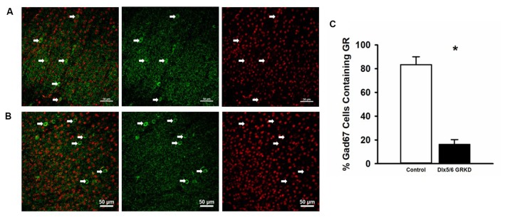
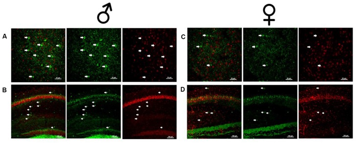
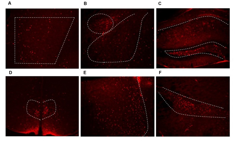
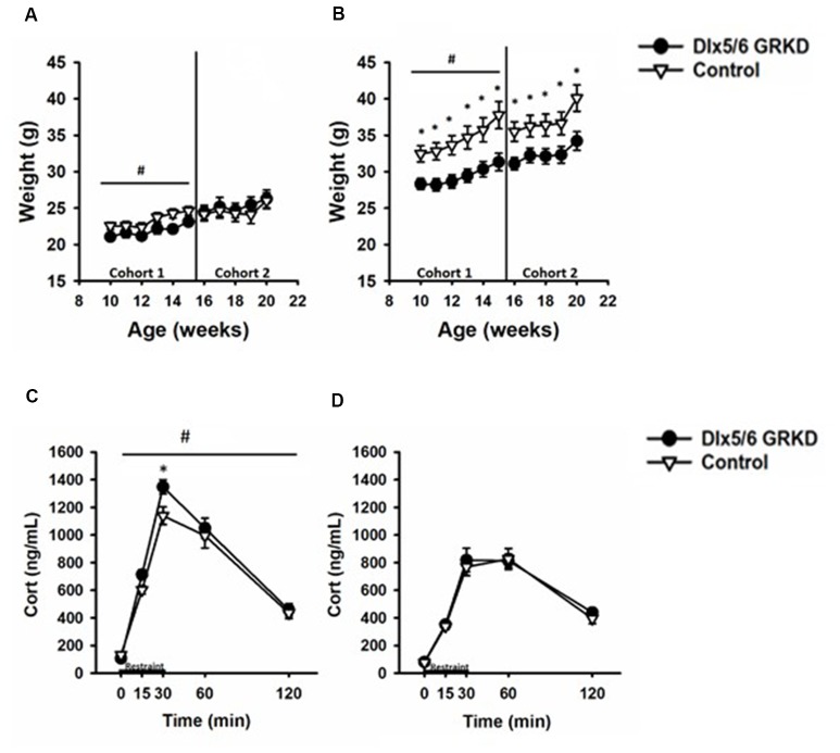
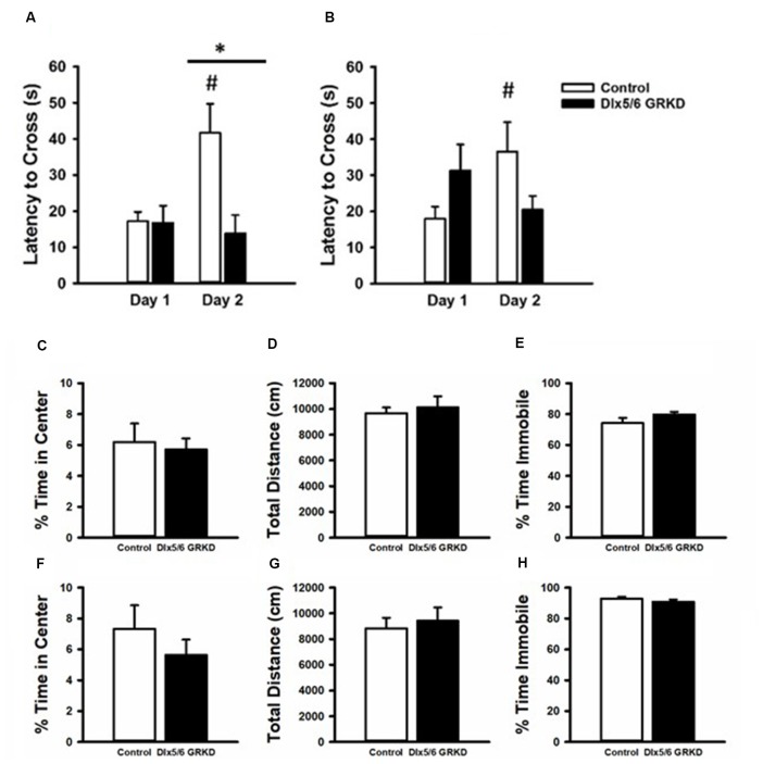
The glucocorticoid receptor (GR) is critically involved in regulation of stress responses [inhibition of the hypothalamic-pituitary-adrenal (HPA) axis], emotional behavior and cognition via interactions with forebrain corticolimbic circuity. Work to date has largely focused on GR actions in forebrain excitatory neurons; however, recent studies suggest a potential role mediated by interneurons. Here, we targeted GR deletion in forebrain GABAergic neurons, including the cortical interneurons, using a Dlx5/6-Cre driver line to test the role of forebrain interneuronal GR in HPA axis regulation and behavior. Our data indicate that GR deletion in GABAergic neurons causes elevated corticosterone stress responsiveness and decreased cross-over latencies in a passive avoidance task in females, but not males. Dlx5/6-Cre driven gene deletion caused loss of GR in interneurons in the prefrontal cortex (PFC) and hippocampus, but also in select diencephalic GABAergic neurons (including the reticular thalamic nucleus and dorsomedial hypothalamus). Our data suggest that GR signaling in interneurons is differentially important in females, which may have implications for GR-directed therapies for stress-related affective disease states.

Keywords: GABAergic; acute stress; glucocorticoid receptor; passive avoidance; sex differences.


Figures

Figure 1
Figure 1
Dlx5/6 GRKD mice show glucocorticoid receptor (GR) knockdown (KD) in most Gad67+ cells. (A) Representative image of a control (Dlx5/6 Cre-; GR f/f) mouse prefrontal cortex (Prelimbic PFC) showing GR staining in Gad67 cells. (B) Representative image of a Dlx5/6 GRKD (Dlx5/6 Cre+; GR f/f) mouse PFC (Prelimbic PFC) showing little to no GR staining in Gad67 cells. (C) Dlx5/6 GRKD mice have significantly fewer cells with definitive GR staining in the Prelimbic. *p ≤ 0.05.
Figure 2
Figure 2
Male and female Dlx5/6 GRKD mice show KD of GR in corticolimbic regions. (A) Male basolateral amygdala. (B) Male hippocampus. (C) Female basolateral amygdala. (D) Female hippocampus.
Figure 3
Figure 3
Dlx5/6 Cre-derived neurons are in areas throughout the forebrain. Representative images of Cre expression in the (A) PFC (B) amygdala (C) hippocampus (D) dorsal medial hypothalamus (DMH) (E) lateral septum (F) zona incerta.
Figure 4
Figure 4
Experimental timeline.
Figure 5
Figure 5
Dlx5/6 GRKD mice gain less weight than controls and female Dlx5/6 GRKD mice have higher peak Corticosterone (Cort) release to acute restraint. (A) Female Dlx5/6 GRKD mice showed a significant interaction between genotype and week in cohort 1, but no individual week was significantly different in post hoc analysis. Cohort 2 females only had a significant effect of time. (B) Male Dlx5/6 GRKD mice weighed less than the controls throughout the study. #p ≤ 0.05 interaction; *p ≤ 0.05 post hoc. (C) Female mice showed an interaction between genotype and time during the acute restraint challenge. Post hoc analysis revealed cort was significantly higher for Female Dlx5/6 GRKD mice at the 30 min point (release point from restraint). (D) Male mice showed no differences in Cort throughout and after the restraint. #p ≤ 0.05 interaction; *p ≤ 0.05 post hoc.
Figure 6
Figure 6
Passive Avoidance test of behavioral inhibition, forced swim test and open field. (A) Female mice showed no difference in latency to cross to the dark compartment on Day 1. While female controls learned and inhibited their behavior, female mice with Dlx 5/6 GRKD had decreased latency to enter the dark compartment compared to controls on day 2. (B) There was no difference in latency from controls in male Dlx 5/6 GRKD mice on day 1 or day 2. (C–H) Females Dlx 5/6 GRKD mice showed no differences in (C) center time or (D) total locomotion in the open field. Female Dlx 5/6 GRKD showed no differences in (E) immobility in the forced swim test. Similarly, male Dlx 5/6 GRKD mice showed no differences in (F) center time or (G) total locomotion in the open field, and immobility (H) in the forced swim test *p ≤ 0.05; #p ≤ 0.05 for comparison of day within group.

References

    1. Allen Z. J., Waclaw R. R., Colbert M. C., Campbell K. (2007). Molecular identity of olfactory bulb interneurons: transcriptional codes of periglomerular neuron subtypes. J. Mol. Histol. 38, 517–525. 10.1007/s10735-007-9115-4 - DOI - PubMed
    1. Bellinger L. L., Bernardis L. L., Brooks S. (1979). The effect of dorsomedial hypothalamic nuclei lesions on body weight regulation. Neuroscience 4, 659–665. 10.1016/0306-4522(79)90142-8 - DOI - PubMed
    1. Bernardis L. L., Bellinger L. L. (1986). Effect of palatable diet on growth, caloric intake and endocrine-metabolic profile in weanling rats with dorsomedial hypothalamic lesions. Appetite 7, 219–230. 10.1016/s0195-6663(86)80027-7 - DOI - PubMed
    1. Bernardis L. L., Bellinger L. L., Goldman J. K., MacKenzie R. (1981). Dorsomedial hypothalamic lesions at weaning and ovariectomy after maturity: somatic and metabolic changes. Physiol. Behav. 26, 91–98. 10.1016/0031-9384(81)90083-4 - DOI - PubMed
    1. Blurton-Jones M., Tuszynski M. H. (2002). Estrogen receptor-beta colocalizes extensively with parvalbumin-labeled inhibitory neurons in the cortex, amygdala, basal forebrain and hippocampal formation of intact and ovariectomized adult rats. J. Comp. Neurol. 452, 276–287. 10.1002/cne.10393 - DOI - PubMed

LinkOut - more resources