Review
. 2018 Dec 21:9:674.
doi: 10.3389/fgene.2018.00674. eCollection 2018.

Centromere and Pericentromere Transcription: Roles and Regulation … in Sickness and in Health

Affiliations
Review

Centromere and Pericentromere Transcription: Roles and Regulation … in Sickness and in Health

Ksenia Smurova et al. Front Genet. .

Abstract

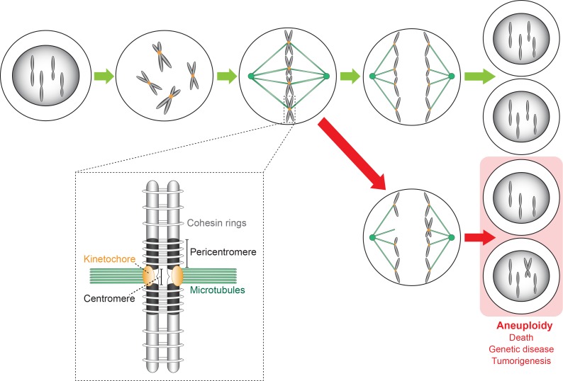
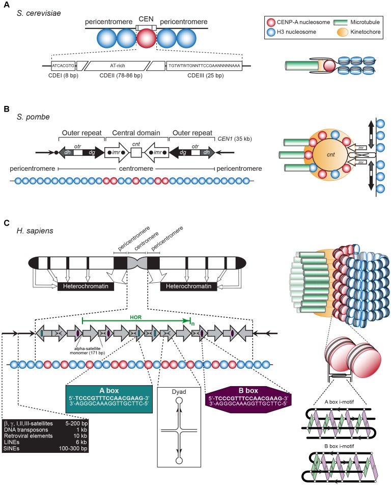
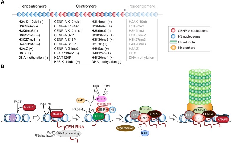
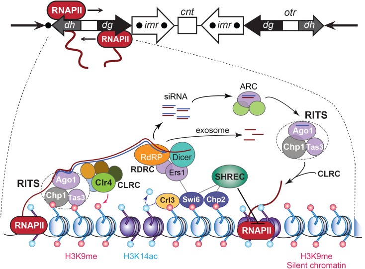
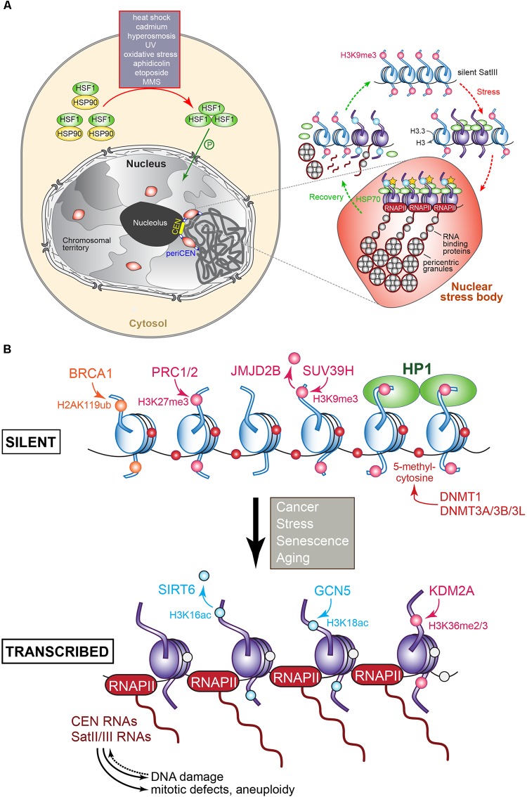
The chromosomal loci known as centromeres (CEN) mediate the equal distribution of the duplicated genome between both daughter cells. Specifically, centromeres recruit a protein complex named the kinetochore, that bi-orients the replicated chromosome pairs to the mitotic or meiotic spindle structure. The paired chromosomes are then separated, and the individual chromosomes segregate in opposite direction along the regressing spindle into each daughter cell. Erroneous kinetochore assembly or activity produces aneuploid cells that contain an abnormal number of chromosomes. Aneuploidy may incite cell death, developmental defects (including genetic syndromes), and cancer (>90% of all cancer cells are aneuploid). While kinetochores and their activities have been preserved through evolution, the CEN DNA sequences have not. Hence, to be recognized as sites for kinetochore assembly, CEN display conserved structural themes. In addition, CEN nucleosomes enclose a CEN-exclusive variant of histone H3, named CENP-A, and carry distinct epigenetic labels on CENP-A and the other CEN histone proteins. Through the cell cycle, CEN are transcribed into non-coding RNAs. After subsequent processing, they become key components of the CEN chromatin by marking the CEN locus and by stably anchoring the CEN-binding kinetochore proteins. CEN transcription is tightly regulated, of low intensity, and essential for differentiation and development. Under- or overexpression of CEN transcripts, as documented for myriad cancers, provoke chromosome missegregation and aneuploidy. CEN are genetically stable and fully competent only when they are insulated from the surrounding, pericentromeric chromatin, which must be silenced. We will review CEN transcription and its contribution to faithful kinetochore function. We will further discuss how pericentromeric chromatin is silenced by RNA processing and transcriptionally repressive chromatin marks. We will report on the transcriptional misregulation of (peri)centromeres during stress, natural aging, and disease and reflect on whether their transcripts can serve as future diagnostic tools and anti-cancer targets in the clinic.

Keywords: centromere; heterochromatin; kinetochore; long non-coding RNA; pericentromere; transcription.


Figures

FIGURE 1
FIGURE 1
Chromosome replication and segregation in a cell undergoing the mitotic cell division cycle. Kinetochores bi-orient the replicated chromosomes (forming sister chromatids) on the metaphase spindle along which they then segregate in opposite directions into the two daughter cells that receive a full complement of the maternal genome (green arrows). Errors made during the segregation process caused by CEN or kinetochore malfunction lead to aneuploid daughter cells (red arrows) carrying an abnormal number of chromosomes. Consequences are cell death, genetic disease (developmental defects), and cancer initiation/progression. The insert shows a more detailed representation of a sister chromosome pair whose chromosomes (original and copy) are linked by cohesion rings. The sister chromosomes are bound to the spindle microtubules via kinetochores that assemble on the CEN sequence of each chromosome.
FIGURE 2
FIGURE 2
(A) Left: The S. cerevisiae point CEN (the consensus CDEI and CDIII sequences are indicated; W = A or T, N = any base). Right: A single CENP-A containing nucleosome is bound to a single microtubule by a single kinetochore (based on Bloom and Costanzo, 2017). (B) Left: The S. pombe regional CEN. Black dots: tRNA clusters. See text for details. Right: A single, looped CEN harboring CENP-A- and histone H3-containing nucleosomes is bound to three microtubules via a single kinetochore (based on McFarlane et al., 2010). (C) Left: A typical human (Homo sapiens) chromosome. White regions: euchromatin, gray region: centromeric chromatin, black regions: heterochromatin. The latter represent the pericentromeres, telomeres, LINEs, SINEs, micro- and macrosatellites, β, γ, I, II, III-satellites, rDNA, and DNA transposons (approximate lengths are indicated in the black box). The gray arrows represent the CEN alpha-satellite monomers, organized in a head-to-tail fashion. HOR, high-order repeat of alpha-satellite monomers (green arrow). A-boxes (dark green) and B-boxes (purple) are indicated, as well as the cruciform configuration of a dyad sequence. Right: Human centromeric chromatin with the CENP-A containing nucleosomes clustered and exposed in amphipathic configuration at its outside is bound by numerous kinetochores to a bundle of microtubule fibers (based on Fukagawa and Earnshaw, 2014). See text for details.
FIGURE 3
FIGURE 3
(A) Epigenetic modifications that mark histones and DNA (cytosines) in the pericentric and centromeric domains. The positive or negative signs indicate whether the modification underlies transcriptional silence or activity, respectively. Modifications of CENP-A required for its deposition or maintenance are also listed. See text for explanations. (B) Schematic outline of transcription-dependent inclusion of histone H3 variant CENP-A at the CEN chromatin, and recruitment of downstream kinetochore components as in vertebrates. See text for details.
FIGURE 4
FIGURE 4
RNA interference-based heterochromatin formation and maintenance at S. pombe CEN. Upper panel: the S. pombe CEN 1, indicating transcription by RNAPII of an outer repeat otr element that flanks the central core of the CEN. Lowe panel: Regulation of the heterochromatic state of CEN sequences that flank the central core domain. The RNA-induced transcriptional silencing (RITS) complex binds to ssRNA transcripts generated from the otr sequence repeats, by siRNA–RNA base pairing interactions and via nucleosomes by localizing to histone H3 methylated at Lys9 (H3K9me). RITS then recruits RDRC/Dicer activity, promotes dsRNA synthesis, the production of siRNAs, and CLRC H3K9 methyltransferase-mediated H3K9 methylation. The Argonaute siRNA chaperone complex (ARC) catalyzes the transfer of the siRNAs from RDRC/Dicer to the RITS complex. The transcript ssRNAs present in the siRNAs become degraded by the exosome. Chromodomain HP1 proteins Swi6 and Chp2 are recruited by the H3K9me mark and silence transcription of the chromatin by localizing the chromatin remodeling Snf2/HDAC repressive complex (SHREC), which inhibits RNAPII activity. Adapted from Holoch and Moazed (2015).
FIGURE 5
FIGURE 5
(A) Regulation of pericentromere SatIII transcription in human cells following exposure to heat and other stresses. In the absence of stress, SatIII repeat sequences are epigenetically marked for silence (H3K9me, pink dot) and exist in a closed transcriptionally inert state (blue nucleosomes). Upon exposure to heat or other stresses, the monomeric HSF1 (shown in green) becomes upregulated, and forms homotrimers that after phosphorylation enter the nucleus. The HSF1 bind to the SatIII sequences and recruit the histone acetyltransferase (HAT) CREB-binding proteins to trigger histone hyperacetylation (yellow stars), which results in active SatIII transcription by RNAPII of one strand. A subset of RNA-binding/processing proteins is recruited to the SatIII transcripts, forming ribonucleoprotein complexes that associate into so-called perichromatin granules, which in turn produce clusters that correspond to a mature nuclear stress body (represented by the red oval structure). To recover from the inflicted stress, heat shock protein HSP70 induces the disassembly of the HSF1 trimers that leave the nuclear stress bodies, along with RNAPII and the HAT. The granules disassemble and the RNA-binding proteins redistribute throughout the nucleoplasm. SatIII transcripts may become processed into smaller fragments possibly by the RNAi machinery to protect and re-establish the heterochromatic state of the pericentromeric region comprising the SatIII repeats, possibly by recruiting epigenetic writing activity resulting in the establishment of the repressive H3K9me signals. Adapted from Biamonti and Vourc’h (2010) and Biamonti (2004). (B) Transcriptional regulation of (peri)centric repeat sequences as identified in various tumors. The epigenetic marks and the enzymes responsible for introducing or removing them at histones or cytosine are indicated. Blue nucleosomes: silent, purple nucleosomes: transcribed. The overproduction of (peri)centric transcripts can induce DNA damage, mitotic defects, genomic instability, and aneuploidy. See text for details.

References

    1. Abudayyeh O. O., Gootenberg J. S., Essletzbichler P., Han S., Joung J., Belanto J. J., et al. (2017). RNA targeting with CRISPR-CAS13. Nature 550 280–284. 10.1038/nature24049 - DOI - PMC - PubMed
    1. Akiyoshi B., Gull K. (2014). Discovery of unconventional kinetochores in kinetoplastids. Cell 156 1247–1258. 10.1016/J.CELL.2014.01.049 - DOI - PMC - PubMed
    1. Albertson D. G., Thomson J. N. (1982). The kinetochores of Caenorhabditis elegans. Chromosoma 86 409–428. 10.1007/BF00292267 - DOI - PubMed
    1. Aldrup-MacDonald M. E., Sullivan B. A. (2014). The past, present, and future of human centromere genomics. Genes 5 33–50. 10.3390/genes5010033 - DOI - PMC - PubMed
    1. Alexandrov I., Kazakov A., Tumeneva I., Shepelev V., Yurov Y. (2001). Alpha-satellite DNA of primates: old and new families. Chromosoma 110 253–266. 10.1007/s004120100146 - DOI - PubMed

LinkOut - more resources