

Retained Intrauterine Device (IUD): Triple Case Report and Review of the Literature
- PMID: 30627466
- PMCID: PMC6304543
- DOI: 10.1155/2018/9362962
Retained Intrauterine Device (IUD): Triple Case Report and Review of the Literature
Abstract
Background: Throughout the world, intrauterine contraceptive devices (IUDs) are a frequently used, reversible, popular contraceptive method. They are usually placed without major complications. Uterine perforation is a rarely observed complication. Migration of the IUD to the pelvic/abdominal cavity or adjacent structures can occur after perforation. We present 3 cases of uterine perforation, possibly due to scarred myometrium associated with a cesarean delivery. We describe 3 perforations with IUDs lodged in the bladder serosa, the posterior cul-de-sac, and tissue adjacent to the cardinal ligament and external iliac artery.
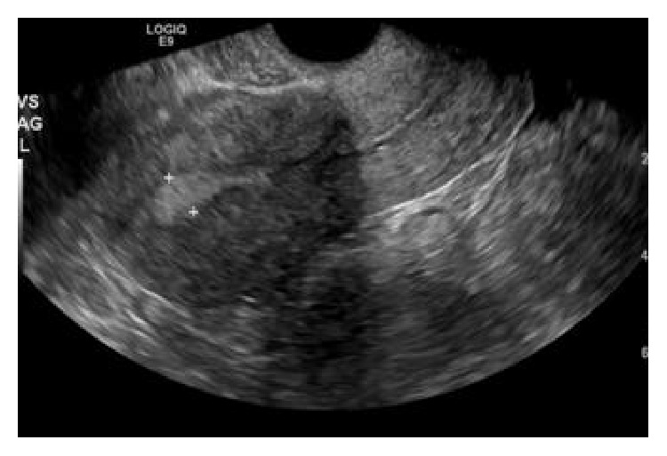
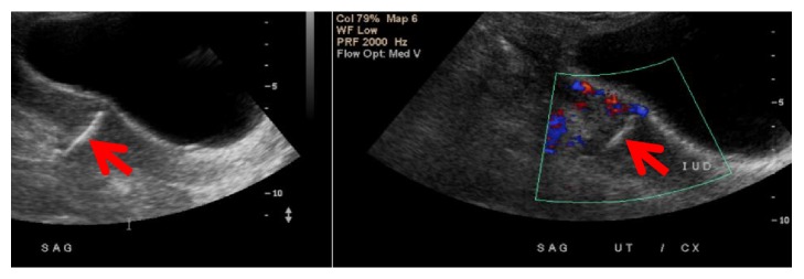
Cases: Case 1. 26-year-old, Gravid 4, Para 2113, nonpregnant female with a history of a cesarean delivery underwent placement of an IUD one year after an elective pregnancy termination, presenting with abdominal pain requesting removal of the IUD. On speculum, although the IUD strings were visualized, the IUD could not be removed. Sonogram imaging identified an empty endometrial cavity with the IUD in posterior cul-de-sac. The IUD was removed via laparoscopy.
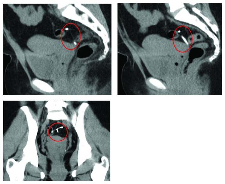
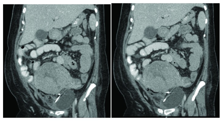
Case 2: 34-year-old Gravida 5, Para 4004, at 27 weeks and 3 days gestation, female with history of two previous cesarean deliveries underwent a third cesarean after spontaneous rupture of membranes with comorbid chorioamnionitis. Reproductive history was significant for placement of an IUD that had not been removed or imaged during obstetrical sonograms. The clinical evaluation revealed that the IUD had been spontaneously expelled. On the fifth operative day, the patient is febrile with CT demonstrating the IUD penetrating the anterior surface of bladder. On cystoscopy the bladder mucosa was intact. The IUD was removed via laparotomy with repair of the bladder, serosa, and muscular layer.
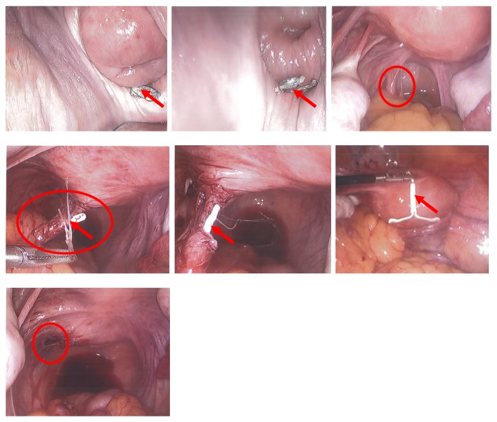
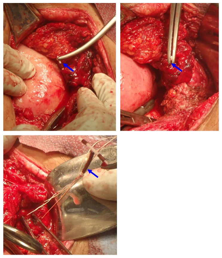
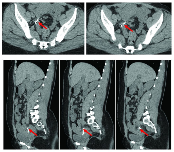
Case 3: 26-year-old, Gravid 4, P3013, nonpregnant female with three previous Cesarean deliveries had an IUD in place. However, with the IUD in situ, the patient conceived and had a spontaneous abortion. After the spontaneous abortion, she presented to clinic to have the IUD removed due to pain that was present since placement. Although the IUD strings were visualized, attempts to remove it were unsuccessful. Imaging identified the IUD outside the uterine cavity. Palpation with a blunt probe laparoscopically revealed a hard object within the adhesion band, close to the cardinal ligament. As per radiology evaluation, IUD was embedded 1cm from the external iliac artery on the right side outside the uterus in the adnexal region. A multidisciplinary procedure with gynecologic-oncologist was scheduled for removal due to the high risk of perioperative bleeding.
Conclusion: Patients in whom uterine perforation and IUD migration are suspected should have appropriate evaluation that includes transvaginal or transabdominal ultrasound or radiographs to confirm the position of the IUD, regardless of whether they are asymptomatic or present with symptoms. It is particularly important in the presence of a scarred uterus that imaging is used to identify the location of a missing IUD. The uterine scar of a cesarean may facilitate migration of the IUD. Cross sectional imaging, such as CT or MRI scan, may be needed to rule out adjacent organ involvement before surgical removal.
Figures
References
-
- CDC. U.S. Selected Practice Recommendations for Contraceptive Use. 2013.
-
- Turner A. Intrauterine device migration. Emergency Medicine Journal. 2016;48(9):417–420. doi: 10.12788/emed.2016.0054. - DOI
-
- Youssouf T., Ibrahim K., Mamadou S., et al. Endoscopic management of migrated intra uterine device in sub-saharan health setting area. Our experience about seventeen cases. Surgical Science. 2018;09(01):17–23. doi: 10.4236/ss.2018.91003. - DOI
Publication types
LinkOut - more resources
Full Text Sources
Research Materials
