Review
. 2018 Dec 27:10:2515841418817490.
doi: 10.1177/2515841418817490. eCollection 2018 Jan-Dec.

Choroideremia: from genetic and clinical phenotyping to gene therapy and future treatments

Affiliations
Review

Choroideremia: from genetic and clinical phenotyping to gene therapy and future treatments

Andreas Mitsios et al. Ther Adv Ophthalmol. .

Abstract

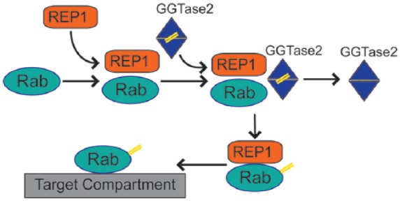
Choroideremia is an X-linked inherited chorioretinal dystrophy leading to blindness by late adulthood. Choroideremia is caused by mutations in the CHM gene which encodes Rab escort protein 1 (REP1), an ubiquitously expressed protein involved in intracellular trafficking and prenylation activity. The exact site of pathogenesis remains unclear but results in degeneration of the photoreceptors, retinal pigment epithelium and choroid. Animal and stem cell models have been used to study the molecular defects in choroideremia and test effectiveness of treatment interventions. Natural history studies of choroideremia have provided additional insight into the clinical phenotype of the condition and prepared the way for clinical trials aiming to investigate the safety and efficacy of suitable therapies. In this review, we provide a summary of the current knowledge on the genetics, pathophysiology, clinical features and therapeutic strategies that might become available for choroideremia in the future, including gene therapy, stem cell treatment and small-molecule drugs with nonsense suppression action.

Keywords: REP1; choroideremia; gene therapy; nonsense suppression therapy; retinal dystrophy; stem cells.


Conflict of interest statement

Conflict of interest statement: The author(s) declared no potential conflicts of interest with respect to the research, authorship and/or publication of this article.

Figures

Figure 1.
Figure 1.
Rab protein prenylation pathway: Rab proteins require lipid modification (prenylation, highlighted as double yellow lines) to associate with membranes and organelles. REP1 mediates the initial step in the process by attaching to Rab proteins and facilitating interaction with geranyl–geranyl transferase 2 enzyme (GGTase2). GGTase 2 prenylates C-terminal cysteine motifs of Rabs and subsequently REP1 delivers modified Rab proteins to target intracellular compartment.
Figure 2.
Figure 2.
Multimodal retinal imaging in a 27-year-old CHM male patient (c.715C>T, p.[Arg239*]). (a) Colour fundus photography shows extensive chorioretinal atrophy with visualisation of scleral vessels and a residual island of retinal tissue in the posterior pole. (b) Fundus autofluorescence of the same eye demonstrates distinct borders between the residual degenerating retinal island and atrophic tissue. (d–g) The green line delineates the level of the optical coherence tomography at the macula, orange and yellow boxes indicate locations for each high-resolution adaptive optics scanning light ophthalmoscopy (AOSLO) image. (c) Optical coherence tomography of the same eye shows disruption of the ellipsoid zone (EZ) (arrowheads) and outer retinal tubulations (ORTs) (arrows). (d–g) AOSLO confocal (D, F) and (e, g) split-detector imaging of the right eye illustrates locations of cone mosaic abrupt disruption at the border of RPE atrophy (yellow arrows in (d) and (e)) in contrast to (f and g) normal cone mosaic in anatomically preserved retinal loci.
Figure 3.
Figure 3.
Colour fundus photography and fundus autofluorescence of (a, c) an unaffected female subject versus (b, d) a CHM female carrier (c.715C>T, p.[Arg239*]). (b) Fundus photograph demonstrates peripapillary changes in CHM female carrier with accompanying peripheral pigmentary and atrophic areas similar to affected male patients. (d) Fundus autofluorescence shows a speckled pattern with intermixed areas of high- and low-density autofluorescence.
Figure 4.
Figure 4.
Optical coherence tomography angiography (OCTA) from the same 27-year-old CHM male patient (c.715C>T, p.[Arg239*]): Macular angiograms (3 × 3) of the superficial and choriocapillaris layers in (a, c) a control male subject versus (b, d) the CHM patient demonstrate decreased vascularity in the choriocapillaris layer of the affected male patient (orange arrows in (d)).

References

    1. Moosajee M, Ramsden SC, Black GC, et al. Clinical utility gene card for: choroideremia. Eur J Hum Genet. Epub ahead of print 21 August 2013. DOI: 10.1038/ejhg.2013.183. - DOI - PMC - PubMed
    1. MacDonald IM, Sereda C, McTaggart K, et al. Choroideremia gene testing. Expert Rev Mol Diagn 2004; 4: 478–484. - PubMed
    1. U.S. National Library of Medicine, National Institutes of Health, Department of Health & Human Services. Choroideremia, https://ghr.nlm.nih.gov/condition/choroideremia#statistics (accessed 2 January 2018).
    1. Sankila EM, Chapelle ADLA, Karna J, et al. Choroideremia: close linkage to DXYS1 and DXYS12 demonstrated by segregation analysis and historical-genealogical evidence. Clin Genet 1987; 31: 315–322. - PubMed
    1. Sankila EM, Tolvanen R, van den Hurk JAJM, et al. Aberrant splicing of the CHM gene is a significant cause of choroideremia. Nat Genet 1992; 1: 109–113. - PubMed