. 2019 May 1;527(7):1278-1289.
doi: 10.1002/cne.24628. Epub 2019 Jan 21.

Selective Cre-mediated gene deletion identifies connexin 43 as the main connexin channel supporting olfactory ensheathing cell networks

Affiliations

Selective Cre-mediated gene deletion identifies connexin 43 as the main connexin channel supporting olfactory ensheathing cell networks

Ana Paula Piantanida et al. J Comp Neurol. .

Abstract

Many functions of glial cells depend on the formation of selective glial networks mediated by gap junctions formed by members of the connexin family. Olfactory ensheathing cells (OECs) are specialized glia associated with olfactory sensory neuron axons. Like other glia, they form selective networks, however, the connexins that support OEC connectivity in vivo have not been identified. We used an in vivo mouse model to selectively delete candidate connexin genes with temporal control from OECs and address the physiological consequences. Using this model, we effectively abolished the expression of connexin 43 (Cx43) in OECs in both juvenile and adult mice. Cx43-deleted OECs exhibited features consistent with the loss of gap junctions including reduced membrane conductance, largely reduced sensitivity to the gap junction blocker meclofenamic acid and loss of dye coupling. This indicates that Cx43, a typically astrocytic connexin, is the main connexin forming functional channels in OECs. Despite these changes in functional properties, the deletion of Cx43 deletion did not alter the density of OECs. The strategy used here may prove useful to delete other candidate genes to better understand the functional roles of OECs in vivo.

Keywords: Cre recombinase; RRID:AB_10000325; RRID:AB_141596; RRID:AB_2110187; RRID:AB_2533973; RRID:AB_2533979; RRID:AB_2576217; RRID:AB_561049; RRID:IMSR_JAX:005975; RRID:IMSR_JAX:007914; RRID:IMSR_JAX:008039; connexin 43; gap junctions; olfactory ensheathing cells; proteolipid protein.


Figures

Figure 1.
Figure 1.. The PLP-Cre line directs Cre expression predominantly in cells in the ONL.
(a) Schematic representation of an OB coronal section indicating the superficial (i) and deep (ii) regions imaged in b. (b) Representative images of coronal sections of the OB from 1-month-old PLP-Cre;Tom mice treated with Tx at 2 weeks of age (P14-P19). Expression of Tom and nuclear stain (DAPI) are shown in red and blue, respectively. Images labeled i-ii correspond to the approximate depths indicated in a. Scale bar: 50 μm. ONL: olfactory nerve layer; GL: glomerular layer; EPL: external plexiform layer; MCL: mitral cell layer; IPL: internal plexiform layer; GCL: granule cell layer. Squared regions at the level of the ONL, GL and MCL/IPL/GCL are magnified in (c). (c) Details of cells labeled by Tom (red) in the different layers of the OB, indicated by arrowheads. The red channel is shown at the right in grayscale. The detail corresponding to the EPL is not shown in (b). Scale bar: 10 μm. (d) Percent of nuclei colocalized with Tom in each OB layer (n=4 mice).
Figure 2.
Figure 2.. The PLP-Cre line efficiently and selectively targets olfactory ensheathing cells.
Representative images of coronal sections of the OB from 1-month-old PLP-Cre;Tom mice treated with Tx at 2 weeks of age (P14-P19). (a) Optical section showing expression of Tom, BLBP and nuclear stain (DAPI) in red, green and blue, respectively. The squared region is shown as combined or separate red and green channels at the right in a magnified scale, indicating Tom-labeled cells with arrowheads. Scale bar: 50 μm (left) and 10 μm (right). ONL: olfactory nerve layer; GL: glomerular layer; EPL: external plexiform layer. (b) Percent of Tom-labeled cells colocalized with the BLBP immunolabeling (left) and percent of BLBPimmunolabeled cells colocalized with Tom expression (right) in the olfactory nerve and glomerular layers (n=3 mice). (c) Confocal stack (maximum projection) of images (7 optical sections at 0.5 μm between sections) showing expression of Tom, GFAP and nuclear stain (DAPI) in red, green and blue, respectively. The squared region is shown as combined or separate red and green channels at the right in a magnified scale. Scale bar: 50 μm μm (left) and 10 μm (right). Cells labeled by Tom expression are indicated with filled arrowheads and GFAP-immunolabeled cells are indicated with open arrowheads. (d) Details of Tom-labeled cells in the deeper layers of the OB showing no colocalization with BLBP in the EPL (top), the mitral cell layer (bottom, open arrowhead) and granule cell layer (bottom, filled arrowhead). Scale bar: 10 μm. Similar results were observed in 3 mice.
Figure 3.
Figure 3.. Cre-Lox mediated deletion of Cx43 in olfactory ensheathing cells without upregulation of Cx30.
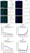
Representative images of coronal sections of the OB from 1-month-old mice immunostained for Cx43 (green) (a-c) or Cx30 (red) (e-g), corresponding to the following genotypes and treatments received at 2 weeks of age (P14-P19): Cx43f/f mouse treated with Tx (a,e), PLP-Cre+/−;Cx43f/f mouse treated with vehicle (b,f) and PLP-Cre+/−;Cx43f/f mouse treated with Tx (c,g). Nuclear stain (DAPI) is shown in blue. The transition from the ONL to the glomerular layer is indicated with a dashed line. Images in grayscale correspond to connexin immunoreactivity alone. Rectangular areas correspond to representative regions of interest used for quantification of intensity profiles and are shown in higher magnification as thresholded images at the bottom of corresponding images. Scale bar: 50 μm. (d,i) Average intensity profiles of Cx43 immunolabeling in the ONL as a function of distance from the glomerular layer outer limit for the experimental groups indicated by the labels, at the juvenile (d) and adult (i) stages. (h,j) Average intensity profiles of Cx30 immunolabel in the ONL as a function of distance to the OB surface for the experimental groups indicated by the labels, at the juvenile (h) and adult (j) stages. Adult mice were treated with Tx at two months of age (P60-P65). ****p<0.0001, post hoc tests after a significant effect of experimental group and distance in a two-way repeated measures ANOVA (n=3–7 juvenile animals and 4–6 adult animals per group). Dotted lines indicate SEM.
Figure 4.
Figure 4.. Cx43 deletion preserves the number of olfactory ensheathing cells.
Representative images of OB sections showing immunoreactivity for BLBP (green) and nuclear stain (DAPI, blue) in control (a) and cCx43KO (b) mice. Higher magnifications of the indicated areas are shown below, with the isolated BLBP channel in grayscale. Filled and open arrowheads point at BLBP+ and BLBP− cells, respectively. Scale bar: 50 μm; inset: 10 μm. (c,d) Average density of BLBP+ cells (c) and nuclei (d) in the ONL of control and cCx43KO mice (not significantly different, Mann-Whitney test, p=0.4069 (BLBP) and p=0.1104 (nuclei), n=5–6 mice per group).
Figure 5.
Figure 5.. Olfactory ensheathing cells from cCx43KO mice are less sensitive to the connexin blocker MFA.
(a) Representative recordings of OEC whole cells currents evoked by a series of voltage steps (−160 to 120 mV, Δ20 mV) before and after adding 100 μM MFA in control (left) and cCx43KO (right) mice. (b) Average I/V curves measured at the time indicated by the arrows in (a). ****p<0.0001, slopes significantly different after post-hoc tests (Tukey) in two-way ANOVA with experimental group and drug as factors and significant interaction (p=0.0032). Data are mean ± SEM. (c,d) Average I/V curves for the MFA-sensitive current in OECs from control and cCx43KO adult (c) and juvenile (d) mice. ***p<0.001, *p=0.0233, significant interaction and main effects in two-way repeated measures ANOVA with voltage and experimental group as factors (adults: n=5–6 cells from 4–5 mice per group; juveniles: n=3–5 cells from 3–4 mice per group). (e) Representative images of OECs filled with lucifer yellow through the recording pipette, obtained at the end of the recording time to qualitatively identify OECs by their characteristic lacunae (arrowheads in the insets). Scale bar: 10 μm.
Figure 6.
Figure 6.. Cx43 mediates gap junction connectivity in olfactory ensheathing cells.
(a) Representative orthogonal projections of image stacks obtained from lucifer yellow (green)-filled OECs from a control mouse (top) and a cCx43KO mouse (bottom). Nuclear stain (DAPI) is shown in blue and the isolated lucifer yellow channel is shown at the right. Scale bar: 20 μm. (b) 3D plots showing the relative distance of neighboring nuclei to the dye-filled cell, centered at the origin of the x-y plane, and the lucifer yellow intensity indicated in the z-axis and color mapped (0 to 255 in arbitrary units), for cells from control (top, n=6 cells from 5 mice) and cCx43KO mice (bottom, n=4 cells from 4 mice). Cells considered below threshold for significant dye staining are indicated in gray.

Similar articles

Cited by

References

    1. Anzini P, Neuberg DH, Schachner M, Nelles E, Willecke K, Zielasek J, … Martini R (1997). Structural abnormalities and deficient maintenance of peripheral nerve myelin in mice lacking the gap junction protein connexin 32. J Neurosci, 17(12), 4545–4551. - PMC - PubMed
    1. Au E, Richter MW, Vincent AJ, Tetzlaff W, Aebersold R, Sage EH, & Roskams AJ (2007). SPARC from olfactory ensheathing cells stimulates Schwann cells to promote neurite outgrowth and enhances spinal cord repair. J Neurosci, 27(27), 7208–7221. doi:10.1523/JNEUROSCI.0509-07.2007 - DOI - PMC - PubMed
    1. Au E, & Roskams A (2003). Olfactory ensheathing cells of the lamina propria in vivo and in vitro. Glia, 41(3), 224–236. - PubMed
    1. Au W, Treloar H, & Greer C (2002). Sublaminar organization of the mouse olfactory bulb nerve layer. J Comp Neurol, 446(1), 68–80. - PubMed
    1. Barnett SC, Thompson RJ, Lakatos A, & Pitts J (2001). Gap junctional communication and connexin expression in cultured olfactory ensheathing cells. J Neurosci Res, 65(6), 520–528. doi:10.1002/jnr.1182 [pii] - DOI - PubMed

Publication types

MeSH terms