. 2019 Mar;43(3):1229-1240.
doi: 10.3892/ijmm.2018.4045. Epub 2018 Dec 31.

Silica nanoparticles induce cardiomyocyte apoptosis via the mitochondrial pathway in rats following intratracheal instillation

Affiliations

Silica nanoparticles induce cardiomyocyte apoptosis via the mitochondrial pathway in rats following intratracheal instillation

Zhongjun Du et al. Int J Mol Med. 2019 Mar.

Abstract

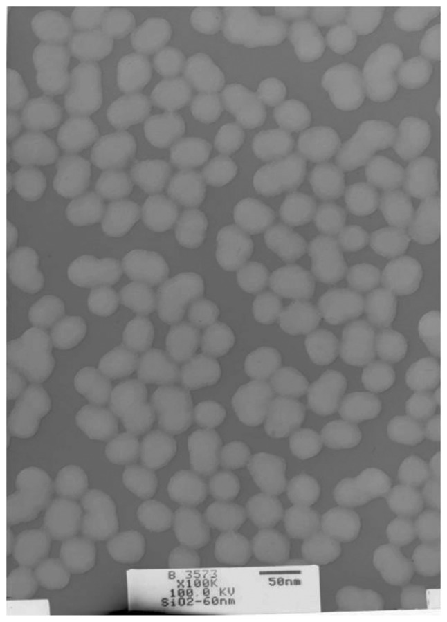
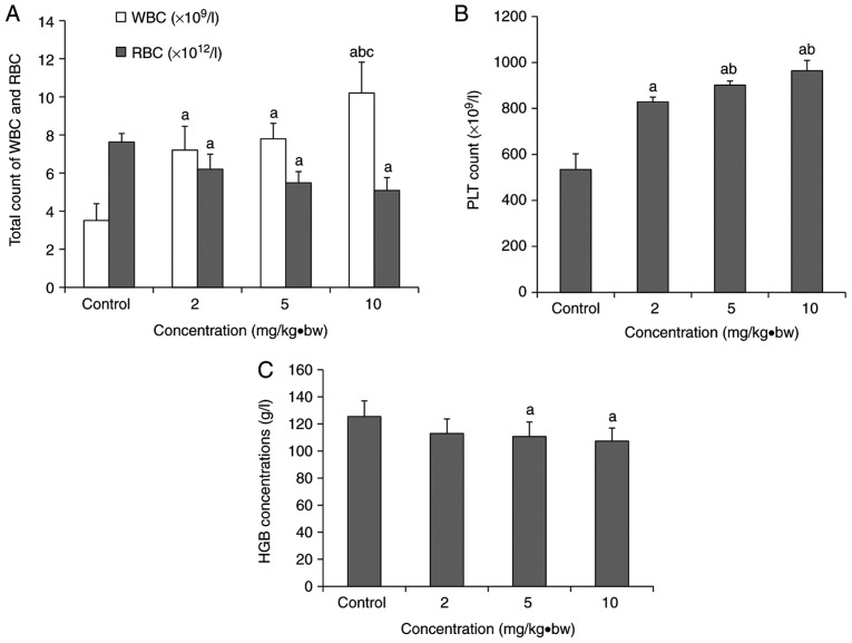
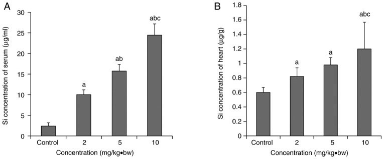
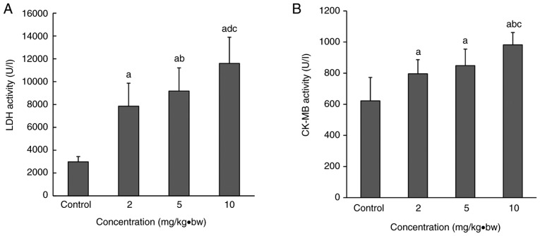
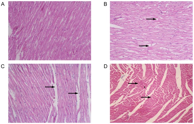
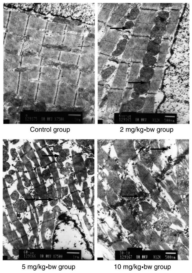
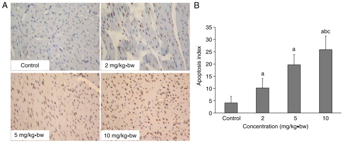
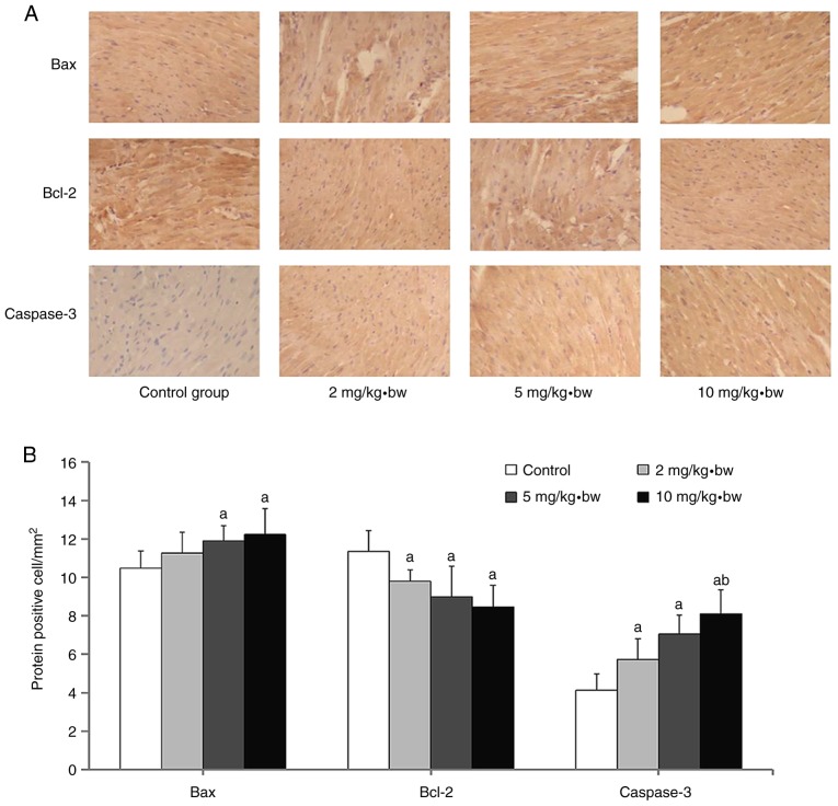
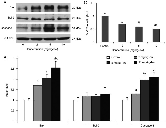
Diseases of the cardiac system caused by silicon dioxide exposure have captured wide public attention. Upon entering the blood circulation, ultrafine particles have the potential to influence cardiomyocytes, leading to myocardial ischemia or even cardiac failure, and the molecular mechanisms remain to be completely elucidated. In this study, the toxicity of ultrafine particles on cardiomyocytes from rats exposed to silica nanoparticles was observed. Rats were randomly divided into a normal saline control group and three exposure groups (2, 5 and 10 mg/kg·body weight) that were intratracheally treated with 60‑nm silica nanoparticles. Alterations in body weight, routine blood factors and myocardial enzymes, histopathological and microstructural alterations, apoptosis and the expression of apoptosis‑associated proteins were assessed at the end of the exposure period. The silicon levels in the heart and serum, and myocardial enzymes in exposed rats were significantly increased in a dose‑dependent manner. In addition, exposure to the silica nanoparticles caused notable histological and ultrastructural alterations in the hearts of these animals. Furthermore, a significant apoptotic effect was observed in the exposure groups. The present data suggest that silica nanoparticles may enter the circulatory system through the lungs, and are distributed to the heart causing cardiovascular injury. Silica nanoparticle‑induced apoptosis via the mitochondrial pathway may serve an important role in observed cardiac damage.


Figures

Figure 1
Figure 1
Transmission electron microscope image of amorphous silica nanoparticles.
Figure 2
Figure 2
Hematological alterations in rats following exposure to silica nanoparticles. (A) The total WBC and RBC counts in each dose group. (B) Platelets count in each dose group. (C) Concentrations of HGB in each dose group (mean ± standard deviation; n=8). aP<0.05 vs. respective control group; bP<0.05 vs. respective 2 mg/kg·bw group; cP<0.05 vs. respective 5 mg/kg·bw group. WBC, white blood cell; RBC red blood cell; PLT, platelets; HGB, hemoglobin; bw, body weight.
Figure 3
Figure 3
Si concentration in the serum and the heart following intratracheal instillation (mean ± standard deviation; n=8). (A) Si concentration in the serum; (B) Si concentration in the heart. aP<0.05 vs. control group; bP<0.05 vs. 2 mg/kg·bw group; cP<0.05 vs. 5 mg/kg·bw group. bw, body weight.
Figure 4
Figure 4
LDH and CK-MB levels in the serum following intratracheal instillation. Serum (A) LDH and (B) CK-MB activities in each dose group were significantly upregulated compared with the control group. A significant increase was also observed in the 10 mg/kg·bw group compared with the 2 and 5 mg/kg·bw groups. Compared with the 2 mg/kg·bw group, LDH activity was significantly upregulated in the 5 mg/kg·bw group. aP<0.05 vs. control group; bP<0.05 vs. 2 mg/kg·bw group; cP<0.05 vs. 5 mg/kg·bw group. CK-MB, creatine kinase-MB; LDH, lactate dehydrogenase; bw, body weight.
Figure 5
Figure 5
Histopathological alterations in cardiomyocytes following intratracheal instillation of silica nanoparticles in rats (magnification, ×200). (A) Control group; (B) 2 mg/kg·bw group; (C) 5 mg/kg·bw group; (D) 10 mg/kg·bw group. In the control group, the cardiomyocytes had a normal morphology. In exposed rats, enlarged intercellular space, fragmented cardiac muscle fibers and obscure boundaries of cardiomyocytes were observed. Histological alterations appeared to increase as the dose increased. Black arrows indicate damaged cardiomyocytes. bw, body weight.
Figure 6
Figure 6
Electron microscopy of cardiomyocytes in rats. In the control group, the cardiomyocytes had a normal morphology. In exposed rats, broken cardiac muscle fibers and dissociated intercalated discs were observed. The mitochondrial membrane was swollen, pyknotic and partially cavitated, while the mitochondria appeared to exhibit vacuolization, and the mitochondrial cristae were broken or absent. Histological alterations appeared to increase as the dose increased. Black arrows indicate intercalated disc dissociation. bw, body weight.
Figure 7
Figure 7
Cardiomyocyte apoptosis in rats following intratracheal instillation of silica nanoparticles. (A) Cardiomyocyte apoptosis and (B) apoptosis indexes of each group following exposure to silica nanoparticles, as assessed by terminal deoxynucleotidyl-transferase-mediated dUTP nick end labelling (magnification, ×200). aP<0.05 vs. control group; bP<0.05 vs. 2 mg/kg·bw group; cP<0.05 vs. 5 mg/kg·bw group. bw, body weight.
Figure 8
Figure 8
Measurement of Bax, Bcl-2 and caspase-3 protein expression in that cardiomyocytes of rats following exposure to silica nanoparticles. (A) Representative immunohistochemical images of Bax, Bcl-2 and caspase-3 proteins are presented (magnification, ×200). (B) Quantification of the number of cells expressing these proteins in the cardiomyocytes of rats. aP<0.05 vs. control group; bP<0.05 vs. 2 mg/kg·bw group. Bax, apoptosis regulator BAX; Bcl-2, apoptosis regulator Bcl-2; bw, body weight.
Figure 9
Figure 9
Bax, Bcl-2 and caspase-3 expression levels, and the Bcl-2/Bax ratio, in rat cardiomyocytes following exposure to silica nanoparticles. (A) Western blotting of Bax, Bcl-2 and caspase-3 at different exposure concentrations; (B) ratio of Bcl-2/Bax at different exposure concentrations; (C) quantification of protein expression at different exposure concentrations. aP<0.05 vs. control group; bP<0.05 vs. 2 mg/kg·bw group; cP<0.05 vs. 5 mg/kg·bw group. Bax, apoptosis regulator BAX; Bcl-2, apoptosis regulator Bcl-2; bw, body weight.

References

    1. Lasfargues M, Stead G, Amjad M, Ding Y, Wen D. In Situ production of copper oxide nanoparticles in a binary molten salt for concentrated solar power plant applications. Materials (Basel) 2017;10:E537. doi: 10.3390/ma10050537. - DOI - PMC - PubMed
    1. Ortelli S, Costa A, Dondi M. TiO2 nanosols applied directly on textiles using different purification treatments. Materials (Basel) 2015;8:7988–7996. doi: 10.3390/ma8115437. - DOI - PMC - PubMed
    1. Oliveira MLS, Navarro OG, Crissien TJ, Tutikian BF, da Boit K, Teixeira EC, Cabello JJ, Agudelo-Castañeda DM, Silva LFO. Coal emissions adverse human health effects associated with ultrafine/nano-particles role and resultant engineering controls. Environ Res. 2017;158:450–455. doi: 10.1016/j.envres.2017.07.002. - DOI - PubMed
    1. Shao H, Mohammed MU, Thomas N, Babazadeh S, Yang S, Shi Q, Shi L. Evaluating excessive burden of depression on health status and health care utilization among patients with hypertension in a nationally representative sample from the medical expenditure panel survey (MEPS 2012) J Nerv Ment Dis. 2017;205:397–404. doi: 10.1097/NMD.0000000000000618. - DOI - PubMed
    1. Hsin YH, Chen CF, Huang S, Shih TS, Lai PS, Chueh PJ. The apoptotic effect of nanosilver is mediated by a ROS- and JNK-dependent mechanism involving the mitochondrial pathway in NIH3T3 cells. Toxicol Lett. 2008;179:130–139. doi: 10.1016/j.toxlet.2008.04.015. - DOI - PubMed