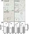
Effects of macrophage depletion on characteristics of cervix remodeling and pregnancy in CD11b-dtr mice
- PMID: 30629144
- PMCID: PMC7302510
- DOI: 10.1093/biolre/ioz002
Effects of macrophage depletion on characteristics of cervix remodeling and pregnancy in CD11b-dtr mice
Abstract
To test the hypothesis that macrophages are essential for remodeling the cervix in preparation for birth, pregnant homozygous CD11b-dtr mice were injected with diphtheria toxin (DT) on days 14 and 16 postbreeding. On day 15 postbreeding, macrophages (F4/80+) were depleted in cervix and kidney, but not in liver, ovary, or other non-reproductive tissues in DT-compared to saline-treated dtr mice or wild-type controls given DT or saline. Within 24 h of DT-treatment, the density of cell nuclei and macrophages declined in cervix stroma in dtr mice versus controls, but birefringence of collagen, as an indication of extracellular cross-linked structure, remained unchanged. Only in the cervix of DT-treated dtr mice was an apoptotic morphology evident in macrophages. DT-treatment did not alter the sparse presence or morphology of neutrophils. By day 18 postbreeding, macrophages repopulated the cervix in DT-treated dtr mice so that the numbers were comparable to that in controls. However, at term, evidence of fetal mortality without cervix ripening occurred in most dtr mice given DT-a possible consequence of treatment effects on placental function. These findings suggest that CD11b+ F4/80+ macrophages are important to sustain pregnancy and are required for processes that remodel the cervix in preparation for parturition.
Keywords: collagen; diphtheria toxin; monocytes; parturition; preterm birth; ripening.
© The Author(s) 2019. Published by Oxford University Press on behalf of Society for the Study of Reproduction.
Figures


Similar articles
-
Immunobiology of Cervix Ripening.Front Immunol. 2020 Jan 24;10:3156. doi: 10.3389/fimmu.2019.03156. eCollection 2019. Front Immunol. 2020. PMID: 32038651 Free PMC article. Review.
-
Progesterone Receptor-Mediated Actions Regulate Remodeling of the Cervix in Preparation for Preterm Parturition.Reprod Sci. 2016 Nov;23(11):1473-1483. doi: 10.1177/1933719116650756. Epub 2016 May 27. Reprod Sci. 2016. PMID: 27233754 Free PMC article.
-
Parturition and recruitment of macrophages in cervix of mice lacking the prostaglandin F receptor.Biol Reprod. 2008 Mar;78(3):438-44. doi: 10.1095/biolreprod.107.063404. Epub 2007 Nov 14. Biol Reprod. 2008. PMID: 18003949 Free PMC article.
-
Monocyte/macrophage suppression in CD11b diphtheria toxin receptor transgenic mice differentially affects atherogenesis and established plaques.Circ Res. 2007 Mar 30;100(6):884-93. doi: 10.1161/01.RES.0000260802.75766.00. Epub 2007 Feb 22. Circ Res. 2007. PMID: 17322176 Free PMC article.
-
Contributions to the dynamics of cervix remodeling prior to term and preterm birth.Biol Reprod. 2017 Jan 1;96(1):13-23. doi: 10.1095/biolreprod.116.142844. Biol Reprod. 2017. PMID: 28395330 Free PMC article. Review.
Cited by
-
Endocrine Disruptor Compounds-A Cause of Impaired Immune Tolerance Driving Inflammatory Disorders of Pregnancy?Front Endocrinol (Lausanne). 2021 Apr 12;12:607539. doi: 10.3389/fendo.2021.607539. eCollection 2021. Front Endocrinol (Lausanne). 2021. PMID: 33912131 Free PMC article. Review.
-
Immunobiology of Cervix Ripening.Front Immunol. 2020 Jan 24;10:3156. doi: 10.3389/fimmu.2019.03156. eCollection 2019. Front Immunol. 2020. PMID: 32038651 Free PMC article. Review.
-
Macrophages exert homeostatic actions in pregnancy to protect against preterm birth and fetal inflammatory injury.JCI Insight. 2021 Oct 8;6(19):e146089. doi: 10.1172/jci.insight.146089. JCI Insight. 2021. PMID: 34622802 Free PMC article.
-
Mixed-Culture Propagation of Uterine-Tissue-Resident Macrophages and Their Expression Properties of Steroidogenic Molecules.Biomedicines. 2023 Mar 22;11(3):985. doi: 10.3390/biomedicines11030985. Biomedicines. 2023. PMID: 36979964 Free PMC article.
-
Is human labor at term an inflammatory condition?†.Biol Reprod. 2023 Jan 14;108(1):23-40. doi: 10.1093/biolre/ioac182. Biol Reprod. 2023. PMID: 36173900 Free PMC article. Review.
References
Publication types
MeSH terms
Substances
Grants and funding
LinkOut - more resources
Full Text Sources
Medical
Molecular Biology Databases
Research Materials

