Fully Automatic Treatment Planning for External-Beam Radiation Therapy of Locally Advanced Cervical Cancer: A Tool for Low-Resource Clinics
- PMID: 30629457
- PMCID: PMC6426517
- DOI: 10.1200/JGO.18.00107
Fully Automatic Treatment Planning for External-Beam Radiation Therapy of Locally Advanced Cervical Cancer: A Tool for Low-Resource Clinics
Abstract
Purpose: The purpose of this study was to validate a fully automatic treatment planning system for conventional radiotherapy of cervical cancer. This system was developed to mitigate staff shortages in low-resource clinics.
Methods: In collaboration with hospitals in South Africa and the United States, we have developed the Radiation Planning Assistant (RPA), which includes algorithms for automating every step of planning: delineating the body contour, detecting the marked isocenter, designing the treatment-beam apertures, and optimizing the beam weights to minimize dose heterogeneity. First, we validated the RPA retrospectively on 150 planning computed tomography (CT) scans. We then tested it remotely on 14 planning CT scans at two South African hospitals. Finally, automatically planned treatment beams were clinically deployed at our institution.
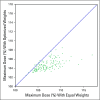
Results: The automatically and manually delineated body contours agreed well (median mean surface distance, 0.6 mm; range, 0.4 to 1.9 mm). The automatically and manually detected marked isocenters agreed well (mean difference, 1.1 mm; range, 0.1 to 2.9 mm). In validating the automatically designed beam apertures, two physicians, one from our institution and one from a South African partner institution, rated 91% and 88% of plans acceptable for treatment, respectively. The use of automatically optimized beam weights reduced the maximum dose significantly (median, -1.9%; P < .001). Of the 14 plans from South Africa, 100% were rated clinically acceptable. Automatically planned treatment beams have been used for 24 patients with cervical cancer by physicians at our institution, with edits as needed, and its use is ongoing.
Conclusion: We found that fully automatic treatment planning is effective for cervical cancer radiotherapy and may provide a reliable option for low-resource clinics. Prospective studies are ongoing in the United States and are planned with partner clinics.
Figures


Similar articles
-
Clinical acceptability of fully automated external beam radiotherapy for cervical cancer with three different beam delivery techniques.Med Phys. 2022 Sep;49(9):5742-5751. doi: 10.1002/mp.15868. Epub 2022 Jul 26. Med Phys. 2022. PMID: 35866442 Free PMC article.
-
Deformable image registration-based contour propagation yields clinically acceptable plans for MRI-based cervical cancer brachytherapy planning.Brachytherapy. 2018 Mar-Apr;17(2):360-367. doi: 10.1016/j.brachy.2017.11.019. Epub 2018 Jan 10. Brachytherapy. 2018. PMID: 29331573
-
Evaluating automatically generated normal tissue contours for safe use in head and neck and cervical cancer treatment planning.J Appl Clin Med Phys. 2024 Jul;25(7):e14338. doi: 10.1002/acm2.14338. Epub 2024 Apr 12. J Appl Clin Med Phys. 2024. PMID: 38610118 Free PMC article.
-
A review of segmentation and deformable registration methods applied to adaptive cervical cancer radiation therapy treatment planning.Artif Intell Med. 2015 Jun;64(2):75-87. doi: 10.1016/j.artmed.2015.04.006. Epub 2015 May 16. Artif Intell Med. 2015. PMID: 26025124 Review.
-
Automated Radiation Treatment Planning for Cervical Cancer.Semin Radiat Oncol. 2020 Oct;30(4):340-347. doi: 10.1016/j.semradonc.2020.05.006. Semin Radiat Oncol. 2020. PMID: 32828389 Free PMC article. Review.
Cited by
-
A Framework for Cervical Cancer Elimination in Low-and-Middle-Income Countries: A Scoping Review and Roadmap for Interventions and Research Priorities.Front Public Health. 2021 Jul 1;9:670032. doi: 10.3389/fpubh.2021.670032. eCollection 2021. Front Public Health. 2021. PMID: 34277540 Free PMC article.
-
Clinical acceptability of fully automated external beam radiotherapy for cervical cancer with three different beam delivery techniques.Med Phys. 2022 Sep;49(9):5742-5751. doi: 10.1002/mp.15868. Epub 2022 Jul 26. Med Phys. 2022. PMID: 35866442 Free PMC article.
-
Access to Radiation Therapy and Related Clinical Outcomes in Patients With Cervical and Breast Cancer Across Sub-Saharan Africa: A Systematic Review.JCO Glob Oncol. 2023 Feb;9:e2200218. doi: 10.1200/GO.22.00218. JCO Glob Oncol. 2023. PMID: 36795990 Free PMC article.
-
Automated Contouring and Planning in Radiation Therapy: What Is 'Clinically Acceptable'?Diagnostics (Basel). 2023 Feb 10;13(4):667. doi: 10.3390/diagnostics13040667. Diagnostics (Basel). 2023. PMID: 36832155 Free PMC article. Review.
-
Artificial Intelligence in Radiotherapy Treatment Planning: Present and Future.Technol Cancer Res Treat. 2019 Jan 1;18:1533033819873922. doi: 10.1177/1533033819873922. Technol Cancer Res Treat. 2019. PMID: 31495281 Free PMC article. Review.
References
-
- Ferlay J, Soerjomataram I, Dikshit R, et al. Cancer incidence and mortality worldwide: Sources, methods and major patterns in GLOBOCAN 2012. Int J Cancer. 2015;136:E359–E386. - PubMed
-
- Bray F. Transitions in human development and the global cancer burden. In: Stewart BW, Wild CP, editors. World Cancer Report 2014. Lyon, France: International Agency for Research on Cancer; 2014. pp. 54–68.
-
- Barton MB, Frommer M, Shafiq J. Role of radiotherapy in cancer control in low-income and middle-income countries. Lancet Oncol. 2006;7:584–595. - PubMed
-
- Barton MB, Jacob S, Shafiq J, et al. Estimating the demand for radiotherapy from the evidence: A review of changes from 2003 to 2012. Radiother Oncol. 2014;112:140–144. - PubMed
-
- Zubizarreta EH, Fidarova E, Healy B, et al. Need for radiotherapy in low and middle income countries – the silent crisis continues. Clin Oncol (R Coll Radiol) 2015;27:107–114. - PubMed

