. 2018 Dec;4(12):e000236.
doi: 10.1099/mgen.0.000236.

Multi-step genomic dissection of a suspected intra-hospital Helicobacter cinaedi outbreak

Affiliations

Multi-step genomic dissection of a suspected intra-hospital Helicobacter cinaedi outbreak

Yasuhiro Gotoh et al. Microb Genom. 2018 Dec.

Abstract

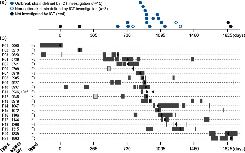
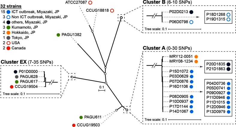
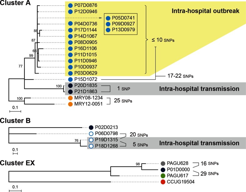
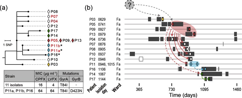
Helicobacter cinaedi is an emerging pathogen causing bacteraemia and cellulitis. Nosocomial transmission of this microbe has been described, but detailed molecular-epidemiological analyses have not been performed. Here, we describe the results of a multi-step genome-wide phylogenetic analysis of a suspected intra-hospital outbreak of H. cinaedi that occurred in a hospital in Japan. The outbreak was recognized by the infectious control team (ICT) of the hospital as a sudden increase in H. cinaedi bacteraemia. ICT defined this outbreak case based on 16S rRNA sequence data and epidemiological information, but were unable to determine the source and route of the infections. We therefore re-investigated this case using whole-genome sequencing (WGS). We first performed a species-wide analysis using publicly available genome sequences to understand the level of genomic diversity of this under-studied species. The clusters identified were then separately analysed using the genome sequence of a representative strain in each cluster as a reference. These analyses provided a high-level phylogenetic resolution of each cluster, identified a confident set of outbreak isolates, and discriminated them from other closely related but distinct clones, which were locally circulating and invaded the hospital during the same period. By considering the epidemiological data, possible strain transmission chains were inferred, which highlighted the role of asymptomatic carriers or environmental contamination. The emergence of a subclone with increased resistance to fluoroquinolones in the outbreak was also recognized. Our results demonstrate the impact of the use of a closely related genome as a reference to maximize the power of WGS.

Keywords: Helicobacter cinaedi; antimicrobial resistance; asymptomatic carrier; intra-hospital outbreak; multi-step phylogenetic analysis.


Conflict of interest statement

The authors declare that there are no conflicts of interest.

Figures

Fig. 1.
Fig. 1.
The 22 H. cinaedi strains isolated in hospital A in Miyazaki used for the re-investigation by WGS analysis of the suspected outbreak. (a) The time of strain isolation (the number of days after the isolation of P01D0000, the earliest isolate in the present strain set) is shown for each strain. (b) The hospitalization periods and admitted wards of the 21 patients (grey bars). Open bars in patients P06 and P12 indicate the admission to other wards. The dates of strain isolation are indicated by closed diamonds.
Fig. 2.
Fig. 2.
The first-step WGS-based phylogenetic analysis. A species-wide analysis of H. cinaedi was performed using 32 strains (22 Miyazaki strains and 10 strains from various other regions) and the complete genome sequence of strain PAGU611 as a reference. Based on the 4154 SNPs identified, an unrooted ML tree was reconstructed using RAxML-NG with the transversion model (TVM). Nodes with ≥80 % bootstrap support based on 1000 bootstrap replicates are indicated by open circles. Three monophyletic clusters identified by TreeGubbins are shown in the boxes outlined with dashed lines, and indistinguishable strains in each cluster are shown in solid boxes. Bars, number of substitutions per site. JP, Japan.
Fig. 3.
Fig. 3.
Phylogenetic analyses of the three clusters identified in the first-step analysis. The three clusters identified in the first-step analysis were separately analysed. SNPs were identified in each cluster using the complete genome sequences of MRY08-1234 (cluster A), P06D0798 (cluster B) and P01D0000 (cluster EX) as references for each cluster to reconstruct ML trees using RAxML-NG with the TPM2 (cluster A) or K80 (clusters B and EX) models. The trees are shown by mid-point rooting. Bootstrap values based on 1000 bootstrap replicates are indicated at nodes. Pairwise SNP distances between isolates are also indicated. The colours of each dot correspond to the strain information indicated in Fig. 2.
Fig. 4.
Fig. 4.
The genetic relationship of the 14 outbreak isolates and their possible transmission routes. (a) The phylogenetic relationships of the 14 outbreak isolates defined in this study are shown as a neighbour-joining tree. The tree was rooted with isolate P15. Diamonds are coloured corresponding to the inferred transmission routes shown in (b). Three isolates that acquired a higher FQ resistance are indicated by asterisks. Five patients with confirmed histories of FQ administration (within 6 months before strain isolation) are indicated in red. The MICs of the 14 isolates to ciprofloxacin (CPFX) and levofloxacin (LVFX) and the mutations (amino acid substitutions) found in GyrA and GyrB in the 14 isolates are also shown. (b) Hospitalization periods of each patient (grey bars), dates of strain isolation (coloured diamonds) and possible strain transmission routes inferred from the phylogenetic relationships of the outbreak isolates are shown. An open bar for patient P12 indicates the admission to a different ward. Dashed arrows indicate the transmissions between patients with no hospitalization period overlap, for which the involvement of ACs or environmental contamination is inferred. Note that a possibility that isolates within a one or two SNP difference (P05/P09/P13, P04, P07, P08, P11, P12 and P14, or some of them) were transmitted from a common source cannot be ruled out (see the main text for more details).

References

    1. Solnick JV, Schauer DB. Emergence of diverse Helicobacter species in the pathogenesis of gastric and enterohepatic diseases. Clin Microbiol Rev. 2001;14:59–97. doi: 10.1128/CMR.14.1.59-97.2001. - DOI - PMC - PubMed
    1. Kawamura Y, Tomida J, Morita Y, Fujii S, Okamoto T, et al. Clinical and bacteriological characteristics of Helicobacter cinaedi infection. J Infect Chemother. 2014;20:517–526. doi: 10.1016/j.jiac.2014.06.007. - DOI - PubMed
    1. Totten PA, Fennell CL, Tenover FC, Wezenberg JM, Perine PL, et al. Campylobacter cinaedi (sp. nov.) and Campylobacter fennelliae (sp. nov.): two new Campylobacter species associated with enteric disease in homosexual men. J Infect Dis. 1985;151:131–139. doi: 10.1093/infdis/151.1.131. - DOI - PubMed
    1. Lasry S, Simon J, Marais A, Pouchot J, Vinceneux P, et al. Helicobacter cinaedi septic arthritis and bacteremia in an immunocompetent patient. Clin Infect Dis. 2000;31:201–202. doi: 10.1086/313930. - DOI - PubMed
    1. Kitamura T, Kawamura Y, Ohkusu K, Masaki T, Iwashita H, et al. Helicobacter cinaedi cellulitis and bacteremia in immunocompetent hosts after orthopedic surgery. J Clin Microbiol. 2007;45:31–38. doi: 10.1128/JCM.01507-06. - DOI - PMC - PubMed

Publication types