. 2019 Jan 10;14(1):e0210660.
doi: 10.1371/journal.pone.0210660. eCollection 2019.

Failure of bumetanide to improve outcome after intracerebral hemorrhage in rat

Affiliations

Failure of bumetanide to improve outcome after intracerebral hemorrhage in rat

Cassandra M Wilkinson et al. PLoS One. .

Abstract

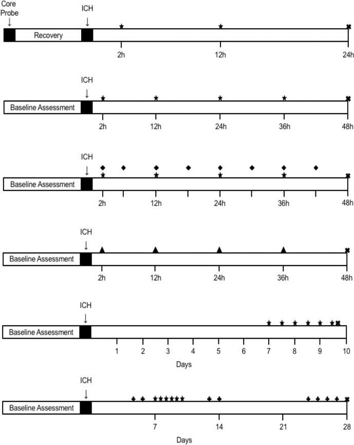
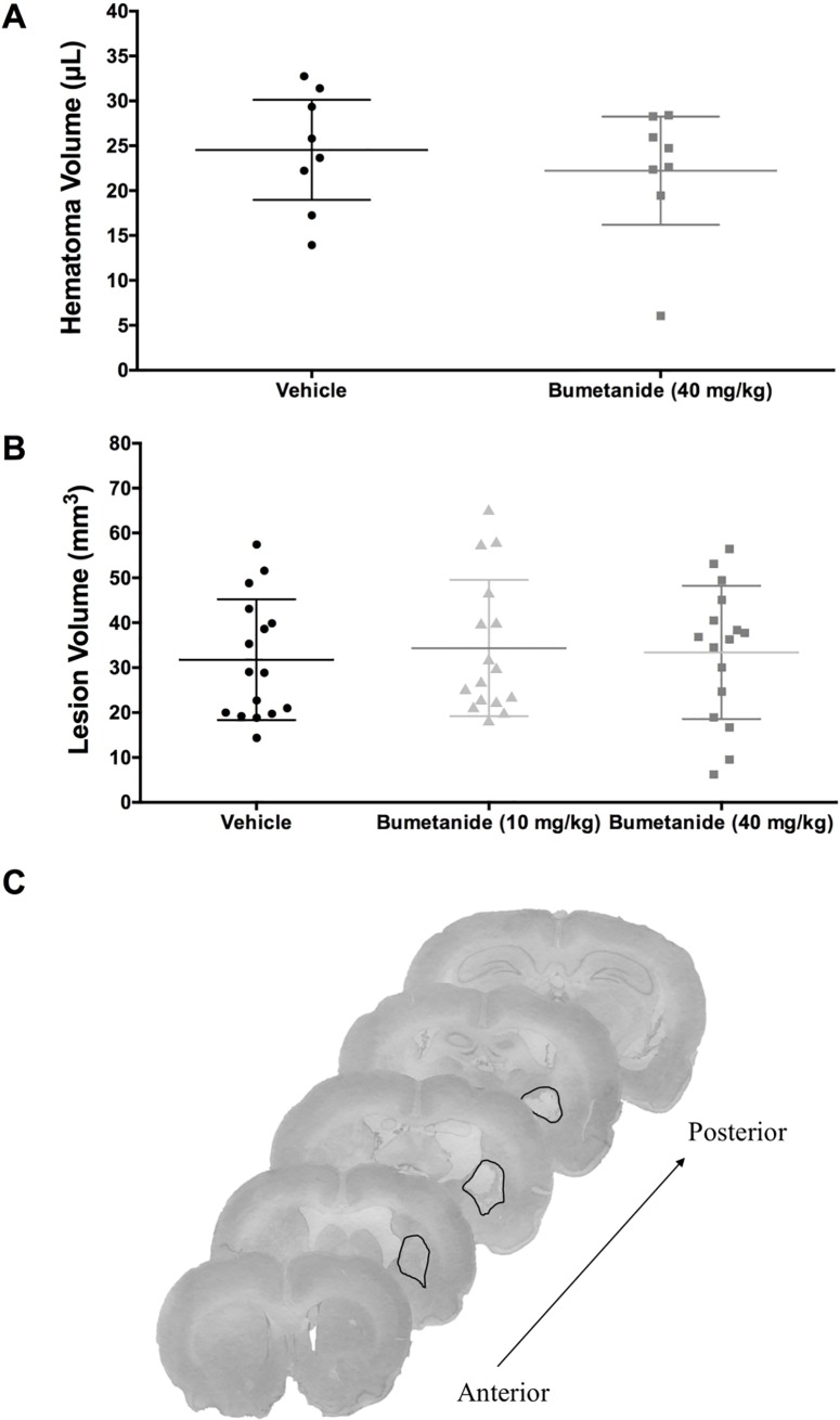
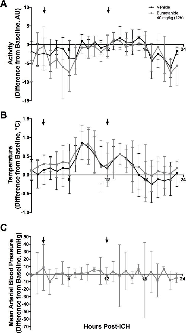
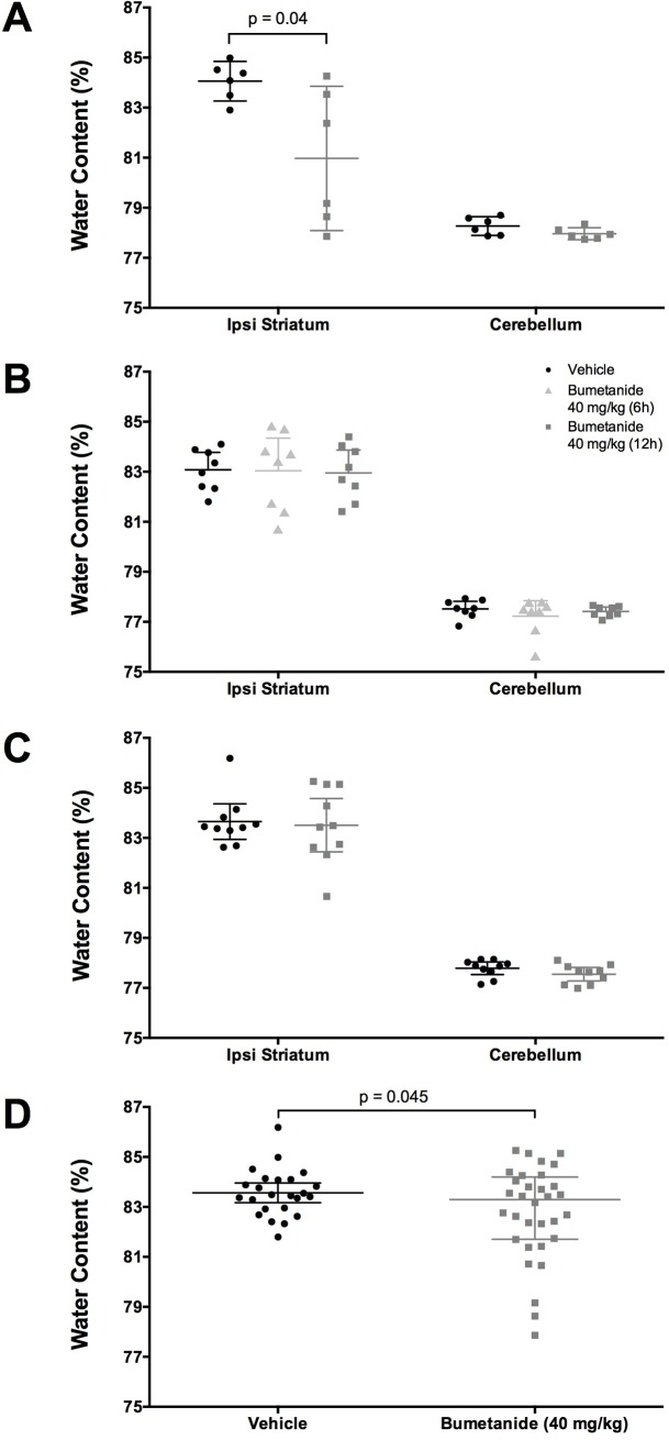
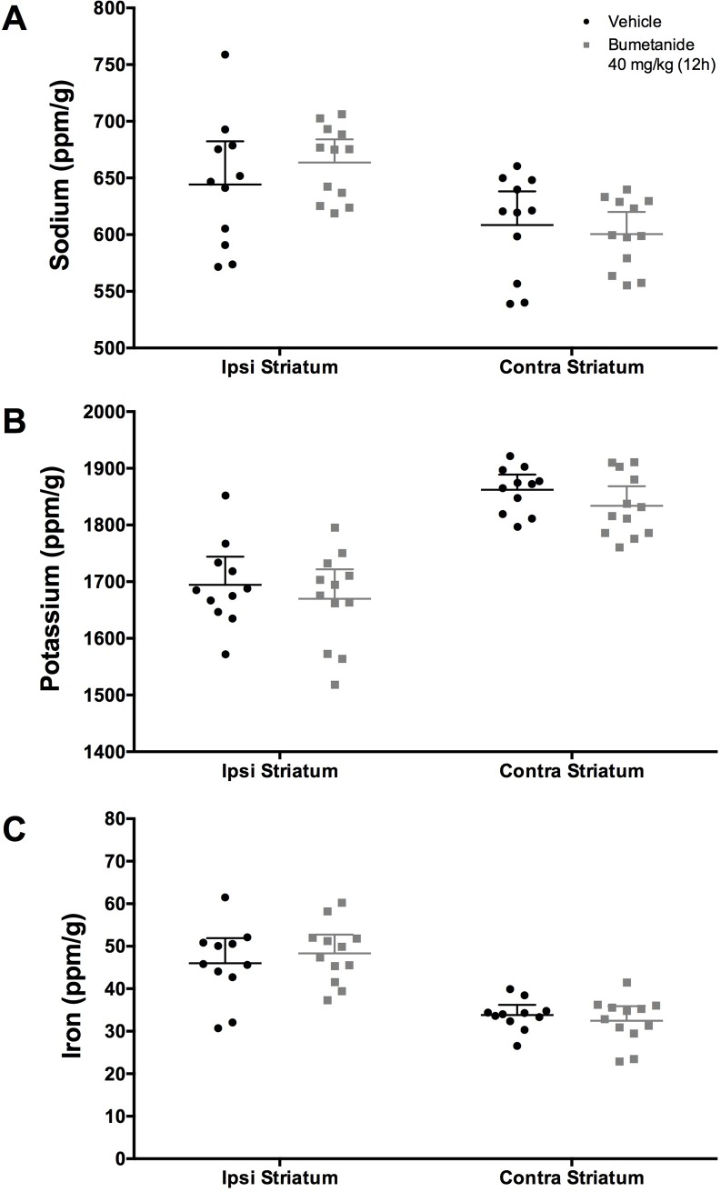
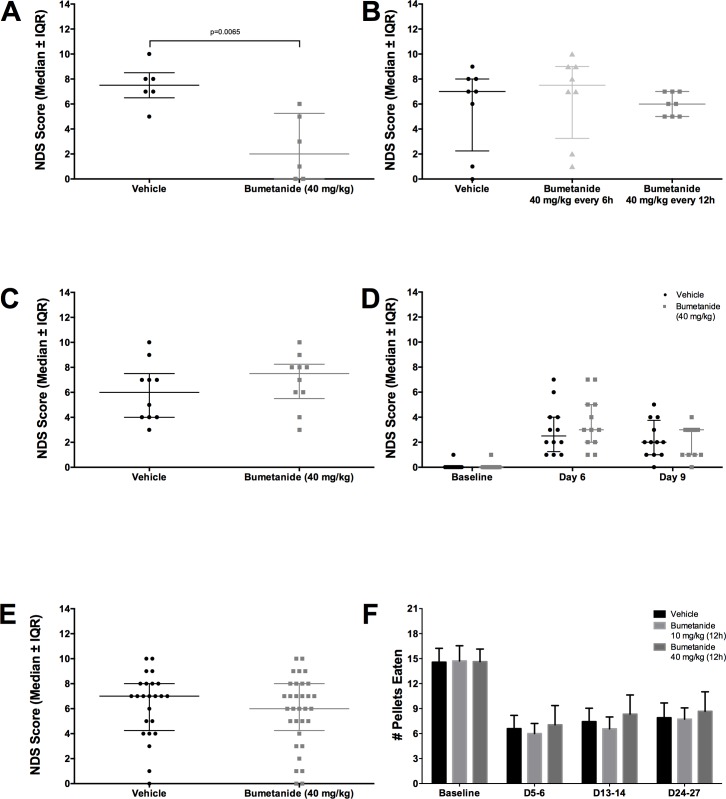
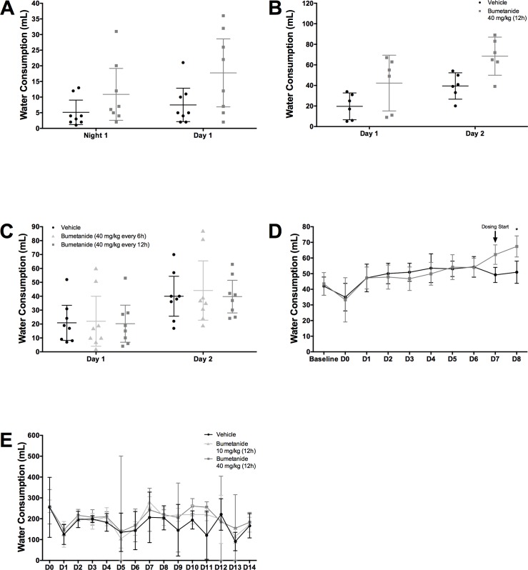
After intracerebral hemorrhage (ICH), brain edema commonly occurs and can cause death. Along with edema, there are significant alterations in the concentrations of key ions such as sodium, potassium, and chloride, which are essential to brain function. NKCC1, a cation-chloride cotransporter, is upregulated after brain damage, such as traumatic injury and ischemic stroke. NKCC1 brings sodium and chloride into the cell, possibly worsening ion dyshomeostasis. Bumetanide, a specific NKCC1 antagonist, blocks the transport of chloride into cells, and thus should attenuate the increases in chloride, which should lessen brain edema and improve neuronal functioning post-ICH, as with other injuries. We used the collagenase model of ICH to test whether bumetanide treatment for three days (vs. vehicle) would improve outcome. We gave bumetanide beginning at two hours or seven days post-ICH and measured behavioural outcome, edema, and brain ion content after treatment. There was some evidence for a minor reduction in edema after early dosing, but this did not improve behaviour or lessen injury. Contrary to our hypothesis, bumetanide did not normalize ion concentrations after late dosing. Bumetanide did not improve behavioural outcome or affect lesion volume. After ICH, bumetanide is safe to use in rats but does not improve functional outcome in the majority of animals.


Conflict of interest statement

The authors have declared that no competing interests exist.

Figures

Fig 1
Fig 1. Experimental Timelines.
(A) Experiment 1. (B) Experiment 2. (C) Experiment 3. (D) Experiment 4. (E) Experiment 5. (F) Experiment 6. ★ denotes oral dose administered on 12-hour interval. ♦ denotes oral dose administered on 6-hour interval. ▲ denotes intraperitoneal (IP) injection on 12-hour interval. ✚ denotes behaviour testing day. ✖ denotes euthanasia.
Fig 2
Fig 2. 24-hour hematoma volume and 28-day lesion volume.
(A) Hematoma volume (μL) data from experiment 1 (n = 8/group) showed that bumetanide did not significantly affect bleeding (P = 0.515). (B) Total tissue loss (mm3), including injury and atrophy, from experiment 6 (n = 16/group) showed that bumetanide did not significantly affect the volume of injury (P = 0.878). (C) Representative histological images at 28 days post-ICH. The black line demarcates the lesion border, and that along with atrophy (e.g., ventriculomegaly) is used to determine tissue loss. This rat, which was in the high dose group, had a total tissue loss of 49.44 mm3. All data presented as mean ± 95% confidence interval.
Fig 3
Fig 3. Temperature, activity, and blood pressure.
(A) Activity (Arbitrary Units, AU) and (B) Temperature (°C) varied after ICH but were not significantly affected by bumetanide (P>0.10, group main effect; P>0.5, interaction effect, n = 8/group). All data presented as mean ± 95% confidence interval. (C) Blood pressure was not significantly affected by bumetanide (P = 0.83).
Fig 4
Fig 4. Brain water content.
(A) Experiment 2 shows that bumetanide reduced brain water content (P = 0.040, n = 6/group). (B) In Experiment 3 (n = 8/group) and (C) Experiment 4 (n = 10/group) shows that bumetanide failed to reduce brain water content (P = 0.275 and P = 0.401 respectively). (D) Pooled brain water content of Experiments 2–4 shows that bumetanide significantly reduced edema (P = 0.045, n = 32 bumetanide-treated, n = 24 vehicle-treated). All data presented as mean ± 95% confidence interval.
Fig 5
Fig 5. Ion concentrations in ipsilateral and contralateral striatum (ppm/g).
Following ICH, (A) Na was increased (P<0.001), (B) K was decreased P<0.001), and (C) Fe was increased (P<0.001) in the ipsilateral striatum as compared to contralateral striatum (n = 11 bumetanide-treated, n = 12 vehicle-treated). Bumetanide did not significantly impact ion concentrations of Na, K, or Fe (P≥ 0.125). All data presented as mean ± 95% confidence interval.
Fig 6
Fig 6. Behavioural assessment.
(A) Data from Experiment 2 showed that bumetanide reduced NDS scores (P = 0.007, n = 6/group). This failed to replicate in (B) Experiments 3 (P = 0.295, n = 8/group), (C) 4 (P = 0.396, n = 10/group), and (D) 5 (P = 0.132, n = 12/group). (E) Pooled NDS Scores of Experiments 2–4 also failed to show an impact of bumetanide on neurological deficits (P = 0.488, n = 32 bumetanide-treated, n = 24 vehicle-treated). (F) Reaching success in Experiment 6 (n = 16/group) showed that ICH significantly reduced reaching ability (P<0.001), which bumetanide failed to improve (P = 0.587). (A-E) presented as median ± interquartile range, (F) presented as mean ± 95% confidence interval.
Fig 7
Fig 7. Hydration.
Animals in the bumetanide groups drank significantly more water in (A) Experiments 1 (P = 0.034, group main effect, n = 8/group), (B) 2 (P = 0.023, group main effect, n = 6/group), (D) 5 (P<0.001 at Day 8, n = 12/group), (E) and 6 (P = 0.029, group main effect, n = 4 cages/group), but not in (C) Experiment 3 (P = 0.932, group main effect, n = 8/group). All data presented as mean ± 95% confidence interval.

References

    1. Ikram MA, Wieberdink RG, Koudstaal PJ. International epidemiology of intracerebral hemorrhage. Curr Atheroscler Rep. 2012; 10.1007/s11883-012-0252-1 - DOI - PMC - PubMed
    1. Williamson MR, Dietrich K, Hackett MJ, Caine S, Nadeau CA, Aziz JR, et al. Rehabilitation Augments Hematoma Clearance and Attenuates Oxidative Injury and Ion Dyshomeostasis After Brain Hemorrhage. Stroke. 2016;48: 195–203. 10.1161/STROKEAHA.116.015404 - DOI - PubMed
    1. Thiex R, Tsirka SE. Brain edema after intracerebral hemorrhage: mechanisms, treatment options, management strategies, and operative indications. Neurosurg Focus. 2007;22: E6 10.3171/foc.2007.22.5.7 - DOI - PubMed
    1. MacLellan CL, Silasi G, Poon CC, Edmundson CL, Buist R, Peeling J, et al. Intracerebral hemorrhage models in rat: comparing collagenase to blood infusion. J Cereb Blood Flow Metab. 2008;28: 516–525. 10.1038/sj.jcbfm.9600548 - DOI - PubMed
    1. Nadeau CA, Dietrich K, Wilkinson CM, Crawford AM, George GN, Nichol HK, et al. Prolonged Blood-Brain Barrier Injury Occurs After Experimental Intracerebral Hemorrhage and Is Not Acutely Associated with Additional Bleeding. Transl Stroke Res. Translational Stroke Research; 2018; 10.1007/s12975-018-0636-9 - DOI - PMC - PubMed

Publication types