. 2019 Jan 9;25(1):39-48.e5.
doi: 10.1016/j.chom.2018.12.004.

Development of a Human Antibody Cocktail that Deploys Multiple Functions to Confer Pan-Ebolavirus Protection

Affiliations

Development of a Human Antibody Cocktail that Deploys Multiple Functions to Confer Pan-Ebolavirus Protection

Anna Z Wec et al. Cell Host Microbe. .

Abstract

Passive administration of monoclonal antibodies (mAbs) is a promising therapeutic approach for Ebola virus disease (EVD). However, all mAbs and mAb cocktails that have entered clinical development are specific for a single member of the Ebolavirus genus, Ebola virus (EBOV), and ineffective against outbreak-causing Bundibugyo virus (BDBV) and Sudan virus (SUDV). Here, we advance MBP134, a cocktail of two broadly neutralizing human mAbs, ADI-15878 from an EVD survivor and ADI-23774 from the same survivor but specificity-matured for SUDV GP binding affinity, as a candidate pan-ebolavirus therapeutic. MBP134 potently neutralized all ebolaviruses and demonstrated greater protective efficacy than ADI-15878 alone in EBOV-challenged guinea pigs. A second-generation cocktail, MBP134AF, engineered to effectively harness natural killer (NK) cells afforded additional improvement relative to its precursor in protective efficacy against EBOV and SUDV in guinea pigs. MBP134AF is an optimized mAb cocktail suitable for evaluation as a pan-ebolavirus therapeutic in nonhuman primates.

Keywords: Ebola glycoprotein; Ebola virus; antibody cocktail; antibody engineering; antiviral drug; broadly neutralizing antibodies; ebolavirus; filovirus; human monoclonal antibodies; immunotherapy.


Figures

Figure 1.
Figure 1.. Selection of ADI-15946 as a candidate cocktail partner for ADI-15878
(A) Hartley guinea pigs were challenged with guinea-pig adapted EBOV (EBOV-GPA) and then treated with single doses of ZMapp, ADI-15878, or vehicle (Dulbecco’s phosphate-buffered saline, DPBS) at three days post-exposure. Survival curves (vehicle vs. test mAb and ZMapp (5 mg) vs. ADI-15878 (5 mg)) were compared by Mantex-Cox test. **, P<0.01. *, P<0.05. (B) Body weights of surviving animals in each treatment group in panel A. Averages±SD (n=6 for ZMapp, n = 3–6 for ADI-15878) are shown. Data are from single cohorts. (C) In silico models of the ADI-15878, ADI-15946, and CA45 Fabs were fitted into negative-stain EM reconstructions of GP:Fab complexes (Wec et al., 2017; Zhao et al., 2017) and are superimposed onto a single EBOV GP structure [PDB ID: 5JQ337] to illustrate the approximate binding footprint and angle of approach of ADI-15946 and CA45 relative to ADI-15878. Light gray and dark gray, GP1 and GP2 subunits, respectively. (D–E) Analysis of competitive binding of candidate IgGs to EBOV GP by biolayer interferometry (BLI). Each GP-bearing probe was sequentially dipped in analyte solutions containing ADI-15878 and then ADI-15878 (D–E, control), CA45 (D), or ADI-15946 (E). Results from a representative experiment are shown.
Figure 2.
Figure 2.. Binding and polyspecificity properties of ADI-15946 and its specificity-matured variant ADI-23774
(A–B) BLI sensorgrams for IgG-SUDV GP interactions with ADI-15946 (A) and ADI-23774 (B). Experimental curves (colored traces) were fit using a 1:1 binding model (black traces). The corresponding flow analyte (GP) concentration is indicated at the right of each curve. (C) Comparison of association (kon) and dissociation (koff) rate constants for IgG interactions with EBOV, BDBV, and SUDV GP. Arrows indicate changes in the values of these constants following ADI-15946 specificity maturation. (D) Polyspecificity scores for candidate mAbs were determined as described previously (Xu et al., 2013). The scores for 137 mAbs in commercial clinical development (Jain et al., 2017) are shown for comparison. Averages±SD (n=3 for ADI-15878 and ADI-15946, n=2 for ADI-23774) from 2–3 independent experiments. See also Figure SI for specificity maturation of ADI-15946 to ADI-23774 and Figure S2 for GP:mAb kinetic binding constants derived from BLI.
Figure 3.
Figure 3.. Neutralizing activity of ADI-23774
Neutralization of rVSVs encoding enhanced green fluorescent protein (eGFP) and bearing uncleaved (A) or cleaved (B) ebolavirus GP proteins (rVSV-GP and rVSV-GPCL, respectively). Virions were preincubated with increasing concentrations of each mAb and then exposed to cells for 12 to 14 hours at 37°C. Infection was measured by automated counting of eGFP+ cells and normalized to infection obtained in the absence of antibody. Averages±SD (n=6–9 in panel A, n=3 in panel B) from 3 independent experiments. (C) Neutralization of authentic filoviruses measured in a microneutralization assay. Virions were preincubated with increasing concentrations of each mAb and then exposed to cells for 48 h at 37°C. Infected cells were immunostained for viral antigen and enumerated by automated fluorescence microscopy. Averages±SD (n=2–4) from two independent experiments.
Figure 4.
Figure 4.. Protective efficacy of ADI-23774 against ebolavirus challenge in mice
(A) BALB/c mice were challenged with mouse-adapted EBOV (EBOV-MA) and then treated with single doses of the indicated mAbs or vehicle (DPBS) at 3 days post-exposure. Survival curves (vehicle vs. test mAb) were compared by Mantex-Cox test. (B) Combined body weights of surviving animals in each treatment group in panel A. Data in panels A–B are from single cohorts. (C) Type 1 IFNα/β R−/− mice were challenged with wild-type SUDV and then treated with two doses of the indicated mAbs or vehicle (DPBS) at 1 and 4 days post-exposure. Survival curves (vehicle vs. test mAb and ADI-15946 vs ADI-23774) were compared by Mantex-Cox test. (D) Combined body weights of surviving animals in each treatment group in panel C. Groups (vehicle vs. test mAb) were compared by two-way ANOVA with repeated measures and Dunnett’s test. Significance values for comparison of body weights on days 6–7 are shown. Data in panels C–D are pooled from two cohorts. ****, P<0.0001. ***, P<0.001. **, P<0.01. *, P<0.05. ns, P>0.05.
Figure 5.
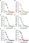
Figure 5.. Neutralizing activity of MBP134 cocktail
(A–F) Neutralization of rVSVs encoding eGFP and bearing GP proteins from EBOV (A), BDBV (B), Taï Forest virus (TAFV) (C), SUDV (D), Reston virus (RESTV) (E), and BOMV (F) was determined as in Figure 3. Averages±SD (n=6) from three independent experiments are shown.
Figure 6.
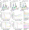
Figure 6.. Protective efficacy of MBP134 in guinea pigs
(A) Hartley guinea pigs were challenged with EBOV-GPA and then treated with single doses of ADI-15878, MBP134 (1:1 mixture of ADI-15878 and ADI-23774), or vehicle (DPBS) at three days post-exposure. Survival curves were compared by Mantex-Cox test. (B) Body weights of surviving animals in each treatment group in panel A. Data are from single cohorts. **, P<0.01. *, P<0.05. ns, P>0.05. See also Table S1 for statistical details on the groups compared in panel A.
Figure 7.
Figure 7.. Development and evaluation of the second-generation MBP134AF cocktail
(A–C) Activation of human natural killer (NK) cells by EBOV GP:IgG complexes. NK cells enriched from the peripheral blood of four different human donors were incubated with complexes between EBOV GP and the indicated IgGs (10 μg/mL) for 5 h at 37°C, and then stained with antibody-fluorophore conjugates specific for the cell-surface markers CD3, CD56, and CD16, followed by intracellular staining for markers of NK cell activation, CD107a (degranulation) (A), IFN-γ (B), and MIP-1β (C). CD3/CD56dim/CD16+ NK cells were analyzed by flow cytometry. Data with cells from all four donors are pooled. HIV-1 glycoprotein-specific mAbs b12 and 2G12 are included as (negative) controls for antigen specificity. EBOV GP-specific mAb c13C6 produced in transgenic Nicotiana benthamiana tobacco plants to bear a highly functional afucosylated/agalactosylated bisected glycan is included as a positive control. Averages±SD (n = 12–14 for all mAbs except b12 and 2G12 (n=6) from 4 independent experiments). The indicated groups were compared by one-way ANOVA and Tukey’s test. (D) Hartley guinea pigs were challenged with EBOV-GPA and then treated with single doses of MBP134, MBP134AF (1:1 mixture of ADI-15878AF and ADI-23774AF), or vehicle (DPBS) at three days post-exposure. (E) Body weights of surviving animals in each treatment group in panel F. Data in panels D–E are from single cohorts. (F–G) Guinea pigs were challenged with SUDV-GPA and then treated with single doses of MBP134AF or vehicle (DPBS) at four (F) or five (G) days post-exposure. Data in panels F–G are from single cohorts. Survival curves in panels D, F were compared by Mantex-Cox test. ****, P<0.0001. **, P<0.01. *, P<0.05. ns, P>0.05. See also Figure S3 for flow-cytometric gating strategies in panels A–C and Table S2 for statistical details on the groups compared in panels D, F.

Comment in

References

    1. Bornholdt ZA, Turner HL, Murin CD, Li W, Sok D, Souders CA, Piper AE, Goff A, Shamblin JD, Wollen SE, et al. (2016). Isolation of potent neutralizing antibodies from a survivor of the 2014 Ebola virus outbreak. Science 351, 1078–1083. - PMC - PubMed
    1. Brannan JM, Froude JW, Prugar LI, Bakken RR, Zak SE, Daye SP, Wilhelmsen CE, and Dye JM (2015). Interferon α/β Receptor-Deficient Mice as a Model for Ebola Virus Disease. J. Infect. Dis 212 Suppl 2, S282–94. - PubMed
    1. Bray M, Davis K, Geisbert T, Schmaljohn C, and Huggins J (1998). A mouse model for evaluation of prophylaxis and therapy of Ebola hemorrhagic fever. J. Infect. Dis 178, 651–661. - PubMed
    1. Burk R, Bollinger L, Johnson JC, Wada J, Radoshitzky SR, Palacios G, Bavari S, Jahrling PB, and Kuhn JH (2016). Neglected filoviruses. FEMS Microbiol. Rev 40, 494–519. - PMC - PubMed
    1. Chao G, Lau WL, Hackel BJ, Sazinsky SL, Lippow SM, and Wittrup KD (2006). Isolating and engineering human antibodies using yeast surface display. Nat. Protoc 1, 755–768. - PubMed

Publication types

MeSH terms