. 2019 Jan 9;25(1):49-58.e5.
doi: 10.1016/j.chom.2018.12.005.

A Two-Antibody Pan-Ebolavirus Cocktail Confers Broad Therapeutic Protection in Ferrets and Nonhuman Primates

Affiliations

A Two-Antibody Pan-Ebolavirus Cocktail Confers Broad Therapeutic Protection in Ferrets and Nonhuman Primates

Zachary A Bornholdt et al. Cell Host Microbe. .

Abstract

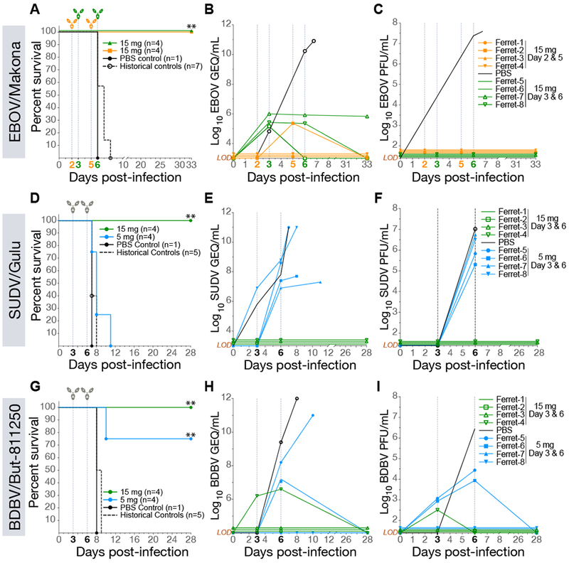
Recent and ongoing outbreaks of Ebola virus disease (EVD) underscore the unpredictable nature of ebolavirus reemergence and the urgent need for antiviral treatments. Unfortunately, available experimental vaccines and immunotherapeutics are specific for a single member of the Ebolavirus genus, Ebola virus (EBOV), and ineffective against other ebolaviruses associated with EVD, including Sudan virus (SUDV) and Bundibugyo virus (BDBV). Here we show that MBP134AF, a pan-ebolavirus therapeutic comprising two broadly neutralizing human antibodies (bNAbs), affords unprecedented effectiveness and potency as a therapeutic countermeasure to antigenically diverse ebolaviruses. MBP134AF could fully protect ferrets against lethal EBOV, SUDV, and BDBV infection, and a single 25-mg/kg dose was sufficient to protect NHPs against all three viruses. The development of MBP134AF provides a successful model for the rapid discovery and translational advancement of immunotherapeutics targeting emerging infectious diseases.

Keywords: ADI-15878; ADI-23774; BDBV; Bundibugyo; EBOV; MBP134; SUDV; Sudan; ebolavirus; pan-ebolavirus.


Conflict of interest statement

DECLARATION OF INTERESTS

A.Z.W., Z.A.B., L.Z., L.M.W., and K.C. are co-inventors on a provisional patent application (No. 62/460,200) assigned to Mapp Biopharmaceutical Inc., Adimab LLC, and Albert Einstein College of Medicine Inc.) describing the development and use of MBP134 and variants thereof for anti-ebolavirus immunotherapy. A.Z.W., E.G., and L.M.W. are employees and shareholders in Adimab LLC. Z.A.B., D.M.A., N.B., O.B., D.H.K., M.H.P., J.V., K.J., and L.Z. are employees and shareholders in Mapp Biopharmaceutical Inc. K.J. and L.Z. are CEO and President of Mapp Biopharmaceutical Inc., respectively.

Figures

Figure 1.
Figure 1.. MBP134AF protects ferrets from lethal EBOV, SUDV and BDBV challenge.
(A) Survival curves for ferrets challenged with EBOV/Makona and treated with 15 mg of MBP134AF on either Day 2 and 5 (orange) or Day 3 and 6 (green) p.i. **, P<0.01. (B) Quantitative RT-PCR measuring average copies of EBOV/Makona genomic equivalents per mL of whole blood (GEQ/mL) from animals treated on day 2 and 5 (orange) and day 3 and 6 (green) p.i. (C) Infectious EBOV/Makona measured via plaque assays (PFU/mL) present in the blood of animals treated on day 2 and 5 (orange) or day 3 and 6 (green). (D) Survival curves for ferrets challenged with SUDV/Gulu and treated with 15-mg (green) or 5-mg (blue) doses of MBP134AF on day 3 and 6 post infection. (E) The average GEQ/mL of SUDV/Gulu present in the blood of animals treated with 15-mg (green) or 5-mg doses (blue). (F) SUDV/Gulu viremia present in the blood of animals treated with 15-mg (green) or 5-mg (blue) doses from panel D. (G) Survival curves for ferrets challenged with BDBV/But-811250 and treated with 15-mg (green) or 5-mg (blue) doses of MBP134AF on day 3 and 6 post infection. (H) The average GEQ/mL of BDBV/But-811250 from animals treated with two 15-mg (green) or 5-mg doses (blue). (I) Viremia (PFU/mL) present in the blood of animals treated with two 15-mg (green) or 5-mg doses (blue) from panel G. LOD = limit of detection
Figure 2.
Figure 2.. A single 25 mg/kg dose of MBP134AF protects rhesus macaques challenged with EBOV/Kikwit.
(A) Survival curves for NHPs challenged with EBOV/Kikwit and treated with a single 25-mg/kg dose of MBP134AF on day 4 (green) p.i or a more conservative two-dose regimen of 50 mg/kg on day 4 and 25 mg/kg on day 7 (orange) post infection. *, P<0.05. (B) The average GEQ/mL of EBOV/Kikwit present in the blood of animals treated with a single dose of MBP134AF (green) or two doses of MBP134AF (orange). All detectable EBOV/Kikwit was eliminated 10 days post treatment. (C) Infectious EBOV/Kikwit (PFU/mL) present in the blood of animals treated with either a single (green) or two-dose course of MBP134AF (orange). Infectious EBOV/Kikwit was no longer detectable by plaque assay by the next bleed of treated animals on day 7 post infection. (D) Clinical scores of animals within the study cohort are shown. Aside from the control animals only NHP-1 and NHP-8 scored for partially or completely refusing their daily nutrition. (E) Body temperatures taken during the course of the study show nine of the ten animals registered an elevated temperature by day 7 p.i. Animals receiving MBP134AF returned to baseline temperature by day 10 p.i. (F) The platelet counts for the cohort show severe thrombocytopenia in the control animals post-infection. By contrast, animals receiving MBP134AF rapidly recovered and displayed little or no signs of thrombocytopenia. Data are represented as mean ± SD. (G) The graphed CRP levels show NHP-3 and NHP-7 suffered from acute inflammation as a result of EVD prior to MBP134AF treatment. (H) The ALT levels from each animal are shown, NHP-3 and NHP-8 demonstrated signs of advanced EVD that were alleviated post-treatment. (I) AST levels for all the challenged animals showed initial signs of liver damage resultant of EVD. Samples from NHP-8 in particular show a significant spike in AST levels that returned to baseline post-treatment. Legend for graphs in top right-hand corner, LOD = limit of detection.
Figure 3.
Figure 3.. Induction of Fc effector function by plant- and CHOKI-produced ADI-23774AF and ADI-15878AF.
(A-C) Markers of antibody-dependent NK cell activation utilizing NK cells from seronegative donors are displayed. The levels of NK cell activation markers CD107a (A), intracellular cytokine IFNγ (B) and MIP-1β (C) are comparatively graphed for ADI-23774 and ADI-15878 produced from transiently expressed HEK293 cells as fucosylated mAbs or as afucosylated mAbs produced from either the plant (-N) or CHOK1-AF (-CHO) cell-based expression platforms. The graphs demonstrate a marked enhancement of Fc-mediated NK cell activation from the presence of an afucosylated glycan in the Fc region of either antibody. Further, both mAbs exceed the Fc effector function potency of c13C6-N, a component of the ZMapp cocktail used here as a positive control. Data are represented as mean ± SD. (D) The phagocytic score or ability of each mAb to facilitate Fc-mediated antibody-dependent neutrophil activation/phagocytosis (ADNP) is shown. Expression in the CHOK1-AF platform greatly enhanced ADNP activity over the other expression platforms likely do to the more uniform heavy chain glycosylation propensity offered by the CHOK1-AF stable pools over transient expression of the antibodies within plants. The formula for calculating the phagocytic score is described in the materials and methods. The anti-human immunodeficiency virus GP120 mAb, 2G12, is utilized here as an irrelevant negative control mAb (Trkola et al., 1996). *, P<0.05. **, P<0.01. ***, P<0.001. ****, P<0.0001.
Figure 4.
Figure 4.. MBP134AF produced from CHOK1-AF cells is therapeutically equivalent to the plant produced MBP134AF.
(A) Hartley guinea pigs were divided into eight treatment groups and given a 5-mg total dose of CHO or plant produced MBP134AF on either Day 4, 5, 6, or 7 post infection. Animals receiving MBP134AF treatment on Day 4 or 5 post infection showed either equivalent and/or significant levels of protection, while all animals treated on day 6 or 7 succumbed to SUDV/Boniface infection with no detectable delay to death when compared to the PBS-treated controls. (B) Weight loss curves from animals treated with CHO or plant produced MBP134AF display equivalent weight change trends. Data are represented as mean ± SD. (C) The viral titers (PFU/mL) in blood samples taken from each animal immediately prior to treatment are graphed. The data suggests MBP134AF produced from either platform equivalently protected animals with <105 PFU/mL of infectious virus, with reduced efficacy in animals that showed viral loads >105 PFU/mL. Data are represented as mean ± SD.
Figure 5.
Figure 5.. A single 25 mg/kg or 7.5 mg/kg dose of MBP134AF protects rhesus macaques challenged with SUDV/Boniface.
(A) Survival curves for NHPs challenged with SUDV/Boniface receiving either PBS (black), a 25-mg/kg dose of MBP134AF (green) or a 7.5-mg/kg (purple) dose of MBP134AF on day 5 p.i. (B) The average GEQ/mL of SUDV/Boniface present in the blood of animals receiving a 25-mg/kg dose of MBP134AF (green), a 7.5-mg/kg dose of MBP134AF (purple) or the PBS controls (black). In the MBP134AF treated groups detectable levels of SUDV/Boniface were eliminated by day 8 post infection (the next bleed post treatment). (C) Infectious SUDV/Boniface (PFU/mL) present in the blood of animals receiving a 25-mg/kg dose of MBP134AF (green) or a 7.5-mg/kg dose of MBP134AF (purple) as well as in PBS controls (black). Infectious SUDV/Boniface was no longer detectable by plaque assay by the next bleed on day 8 p.i. (D) All of the MBP134AF treated animals in the cohort had clinical scores on day 5 p.i. prior to receiving treatment. Animals dosed with MBP134AF no longer registered a clinical score by day 14 p.i., while the surviving controls continued scoring out to day 21 p.i. (E) Body temperatures taken from the blinded cohort showed all the animals registered an elevated temperature by day 5 p.i., prior to receiving MBP134AF. (F) The platelet counts for the cohort show declining counts p.i. with the MBP134AF treated animals rapidly recovering and the PBS controls displaying severe thrombocytopenia. Data are represented as mean ± SD. (G) The ALT levels from each animal are shown; the control animals all display elevated levels from baseline post infection. In contrast, all of the animals receiving MBP134AF show little to no signs of EVD. (H) AST levels for all the MBP134AF treated animals show little to no increase, while the control animals all show highly elevated AST levels by day 10 p.i. (I) ALP levels are graphed for each animal in the cohort, no sign of EVD induced ALP levels are present in any of the MBP134AF treated animals. In contrast, all of the control animals that received PBS display elevated ALP levels resulting from EVD. Legend for graphs in top right-hand corner, LOD = limit of detection.
Figure 6.
Figure 6.. A single 25 mg/kg dose of MBP134AF protects cynomolgus macaques challenged with BDBV/But-811250.
(A) Survival curves for NHPs challenged with BDBV/But-811250 and treated with a single 25-mg/kg dose of MBP134AF on day 7 post infection (green or pink) or PBS (black). **, P<0.01. (B) The average GEQ/mL of BDBV/But-811250 present in the blood of animals treated with a single dose of MBP134AF (green or pink) or PBS (black). Serum samples taken from NHP-4 and NHP-6 on days 20 and 28 tested negative for viral genetic material. (C) Infectious BDBV/But-811250 (PFU/mL) present in the blood of animals treated with MBP134AF (green or pink) or PBS (black). Infectious BDBV was no longer detectable by plaque assay by the next bleed on day 10 post infection in the surviving animals. (D) Clinical scores show NHP-5 and NHP-6 registering scores from refusing nutrition, having a hunched posture and displaying petechiation over 10% of their bodies prior to receiving MBP134AF on day 7 p.i. While NHP-6 cleared clinical signs of infection by day 12 p.i., NHP-5 failed to recover and succumbed to infection on 13 p.i. (E) Body temperatures from all the animals showed the majority registered an elevated temperature by day 7 p.i. prior to receiving MBP134AF. (F) The platelet counts for the cohort show thrombocytopenia occurring in all the animals by day 7 p.i. prior to receiving MBP134AF. All the MBP134AF treated animals (excluding NHP-5, pink) cleared signs of thrombocytopenia by day 14 p.i., seven days post treatment. (G) ALP levels are graphed for each animal in the cohort. (H) ALT levels from each animal are shown. Notably NHP-5 (pink) displayed advanced signs of EVD prior to treatment. (I) AST levels for the challenged animals show varying signs of liver damage resultant of EVD. Samples from NHP-5 (pink) in particular show a significant spike in AST levels suggesting a severe onset of disease beyond that of the other animals in the cohort, a possible explanation of NHP-5’s succumbing to EVD despite receiving the MBP134AF treatment course. Legend for graphs in top right-hand corner, LOD = limit of detection.

Comment in

References

    1. Bornholdt ZA, Turner HL, Murin CD, Li W, Sok D, Souders CA, Piper AE, Goff A, Shamblin JD, Wollen SE, et al. (2016). Isolation of potent neutralizing antibodies from a survivor of the 2014 Ebola virus outbreak. Science 351, 1078–1083. - PMC - PubMed
    1. Burk R, Bollinger L, Johnson JC, Wada J, Radoshitzky SR, Palacios G, Bavari S, Jahrling PB, and Kuhn JH (2016). Neglected filoviruses. FEMS Microbiol Rev. - PMC - PubMed
    1. Corti D, Misasi J, Mulangu S, Stanley DA, Kanekiyo M, Wollen S, Ploquin A, Doria-Rose NA, Staupe RP, Bailey M, et al. (2016). Protective monotherapy against lethal Ebola virus infection by a potently neutralizing antibody. Science. - PubMed
    1. Cross RW, Mire CE, Borisevich V, Geisbert JB, Fenton KA, and Geisbert TW (2016). The Domestic Ferret (Mustela putorius furo) as a Lethal Infection Model for 3 Species of Ebolavirus. J Infect Dis 214, 565–569. - PMC - PubMed
    1. Cross RW, Speranza E, Borisevich V, Widen SG, Wood TG, Shim RS, Adams RD, Gerhardt DM, Bennett RS, Honko AN, et al. (2018). Comparative Transcriptomics in Ebola Makona-Infected Ferrets, Nonhuman Primates, and Humans. J Infect Dis 10.1093/infdis/jiy455. - DOI - PMC - PubMed

Publication types

MeSH terms