

Regional Anesthesia for Dentistry and Orofacial Surgery in Rhesus Macaques (Macaca mulatta)
- PMID: 30630557
- PMCID: PMC6433348
- DOI: 10.30802/AALAS-JAALAS-18-000068
Regional Anesthesia for Dentistry and Orofacial Surgery in Rhesus Macaques (Macaca mulatta)
Abstract
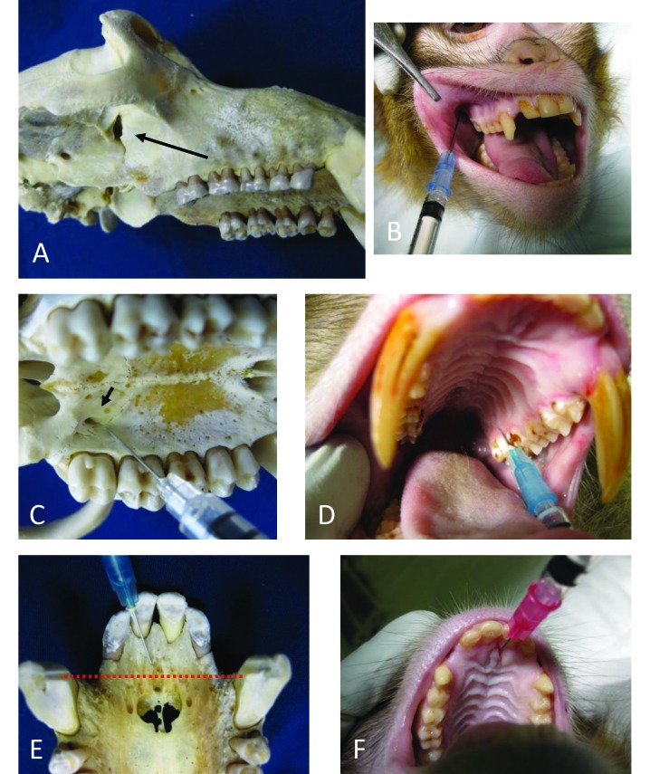
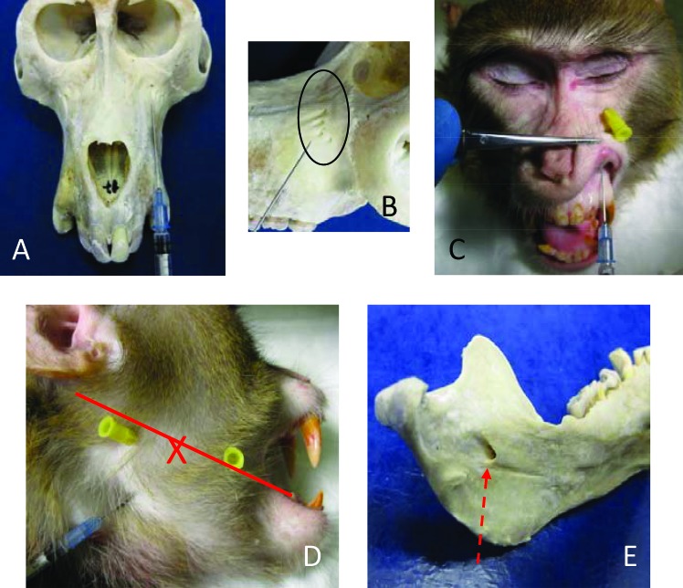
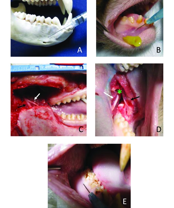
Regional anesthesia is a commonly used adjunct to orofacial dental and surgical procedures in companion animals and humans. However, appropriate techniques for anesthetizing branches of the mandibular and maxillary nerves have not been described for rhesus monkeys. Skulls of 3 adult rhesus monkeys were examined to identify relevant foramina, establish appropriate landmarks for injection, and estimate injection angles and depth. Cadaver heads of 7 adult rhesus monkeys (4 male, 3 female) were then injected with thiazine dye to demonstrate correct placement of solution to immerse specific branches of the mandibular and maxillary nerves. Different volumes of dye were injected on each side of each head to visualize area of diffusion, and to estimate the minimum volume needed to saturate the area of interest. After injection, the heads were dissected to expose the relevant nerves and skull foramina. We describe techniques for blocking the maxillary nerve as well as its branches: the greater palatine nerve, nasopalatine nerve, and infraorbital nerve. We also describe techniques for blocking branches of the mandibular nerve: inferior alveolar nerve, mental (or incisive) nerve, lingual nerve, and long buccal nerve. Local anesthesia for the mandibular and maxillary nerves can be accomplished in rhesus macaques and is a practical and efficient way to maximize animal welfare during potentially painful orofacial procedures.
Figures
Similar articles
-
Variant Inferior Alveolar Nerves and Implications for Local Anesthesia.Anesth Prog. 2016 Summer;63(2):84-90. doi: 10.2344/0003-3006-63.2.84. Anesth Prog. 2016. PMID: 27269666 Free PMC article.
-
A rare case of the auriculotemporal and inferior alveolar nerves communication.Surg Radiol Anat. 2024 Feb;46(2):191-194. doi: 10.1007/s00276-023-03283-9. Epub 2023 Dec 27. Surg Radiol Anat. 2024. PMID: 38151551
-
Variability in the number of infraorbital foramina in rhesus macaques (Macaca mulatta) and cynomolgus macaques (Macaca fascicularis).Anat Rec (Hoboken). 2021 Apr;304(4):818-831. doi: 10.1002/ar.24478. Epub 2020 Jul 11. Anat Rec (Hoboken). 2021. PMID: 32558307
-
A review of the intraosseous course of the nerves of the mandible.J Oral Implantol. 1991;17(4):394-403. J Oral Implantol. 1991. PMID: 1813647 Review.
-
Applied anatomy of the pterygomandibular space: improving the success of inferior alveolar nerve blocks.Aust Dent J. 2011 Jun;56(2):112-21. doi: 10.1111/j.1834-7819.2011.01312.x. Aust Dent J. 2011. PMID: 21623801 Review.
Cited by
-
Selection of points for regional anesthesia of the trigeminal nerve and its branches in red pandas (Ailurus fulgens fulgens).Vet Res Commun. 2025 Apr 25;49(3):179. doi: 10.1007/s11259-025-10746-4. Vet Res Commun. 2025. PMID: 40278969
-
Topography of cranial foramina and anaesthesia techniques of cranial nerves in selected species of primates (Cebidae, Cercopithecidae, Lemuridae) - part I - osteology.BMC Vet Res. 2023 Aug 12;19(1):122. doi: 10.1186/s12917-023-03680-7. BMC Vet Res. 2023. PMID: 37573315 Free PMC article.
References
-
- Alzarea BK. 2014. Selection of animal models in dentistry: State of art, review article. J Anim Vet Adv 13:1080–1085.
-
- Christensen K. 1961. The cranial nerves, p 290–306. In: Hartman CG, Straus WL, The Anatomy of the Rhesus Monkey, New York (NY) Hafner Publishing Co.
MeSH terms
LinkOut - more resources
Full Text Sources
Medical
