. 2019 Jan 10;10(1):23.
doi: 10.1038/s41419-018-1255-9.

Lamin B is a target for selective nuclear PQC by BAG3: implication for nuclear envelopathies

Affiliations

Lamin B is a target for selective nuclear PQC by BAG3: implication for nuclear envelopathies

Manish K Gupta et al. Cell Death Dis. .

Abstract

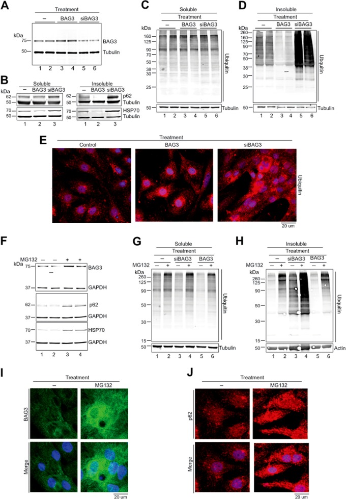
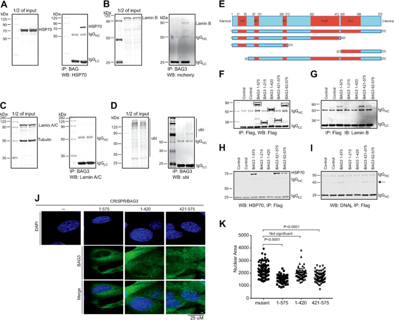
Nuclear envelopathies are recognized genetic disorders affecting individuals with mutations in their genes encoding members of the lamin family of nuclear envelope proteins that are responsible for maintaining the architectural structure of the nucleus. Irregularity in shape and size of the nuclei, nuclear membrane rupture, and appearance of micronuclei in the cytoplasm are among the pathological features of the syndrome. Here, we demonstrate that Bcl2-associated anthanogene-3 (BAG3), a stress-induced co-chaperone protein that by association with heat-shock protein 70 (HSP70) participates in regulation of autophagy, plays a critical role in the integrity of the nuclear membrane in cardiomyocytes. Cells subjected to proteotoxic stress or BAG3 downregulation show perinuclear accumulation of the aberrant ubiquitinated proteins that are often associated with the appearance of misshapen, enlarged, and elongated nuclei. There were dense accumulations of lamin B in the perinuclear area and distribution of lamin B-positive micronuclei in the cytoplasmic space, indicative of nuclear envelope rupture. Overexpression of BAG3 in cells under proteotoxic stress ameliorated pathological nuclear morphology and reduced cytoplasmic distribution of the micronuclei particles. Subcellular co-localization and co-immunoprecipitation demonstrated interaction of lamin B with the BAG domain of BAG3 and HSP70, suggesting the importance of BAG3 in the selective clearance of a surplus of aggregated lamin B that is generated during stress conditions. Our findings define a novel role for BAG3 in nuclear protein quality control and suggest an alternative pathogenetic pathway that contributes to the development of nuclear envelopathies.


Conflict of interest statement

The authors declare no competing interests.

Figures

Fig. 1
Fig. 1. BAG3 is important for maintaining cellular protein quality control.
a Reduced expression of BAG3 disrupts cellular proteostasis and causes accumulation of aggregated proteins. Cardiomyocytes were transduced with Ad-GFP, Ad-BAG3, and Ad-siBAG3 for 48 h. Expression of BAG3 was determined in the total cellular fraction. b–d Soluble and insoluble fractions were prepared from the transfected cardiomyocytes and expression of p62, HSP70, and ubiquitin was determined by western blotting. Expression was normalized with Tubulin. e Representative images show that knockdown of BAG3 causes accumulation of ubiquitin-positive cellular aggregates in cardiomyocytes. Cardiomyocytes were transduced with the respective adenovirus and cells were fixed with 4% PFA. Fixed cells were subsequently stained with ubiquitin antibody (red) and nuclei were stained with DAPI. f–h Inhibition of proteasomal function upregulates BAG3 expression in the cardiomyocytes. Cardiomyocytes were treated with MG132 (20 μM) for 6 h and expression of BAG3 was evaluated by western blot. Expression of p62, HSP70, and ubiquitin was assessed by western blot in cardiomyocytes treated with MG132 for 6 h. g–h Knockdown of BAG3 increases accumulation of ubiquitinated proteins in the insoluble fraction. NRVCs were transduced with adenovirus for 48 h and treated with MG132 before protein isolation. Western blot was done with ubiquitin antibody in the soluble and insoluble fractions. i Representative microscopy images show that inhibition of proteasomal function causes upregulation of BAG3 protein in cardiomyocytes. Cardiomyocytes were treated with MG132 for 6 h and cells were fixed with 4% PFA. Fixed cells were then stained with BAG3 antibody (green) and nuclei were stained with DAPI (blue). j Fixed cells were stained with p62 antibody (red) and nuclei were stained with DAPI
Fig. 2
Fig. 2. BAG3 is important for removal of nuclear protein during proteotoxic stress.
a, b Western blots show that due to proteasomal inhibition, expression of BAG3 and lamin B was upregulated in the cytosolic fraction as well as in the nuclear fraction. NRVCs were treated with the proteasomal inhibitor MG132 (20 µM) for 6 h and cells were fractionated into cytosolic and nuclear fractions. Expression of BAG3 and lamin B, HSP70, and p62 was determined by western blot in the cytosolic as well as in nuclear fraction by BAG3, lamin B, HSP70, and p62 antibodies, respectively. Expression of cytosolic and nuclear proteins was normalized with GAPDH and lamin A/C expression, respectively. Graph shows the quantification of BAG3, HSP70, p62, and lamin B expression in the cytosol as well as in the nuclear fraction. c, d) Proteasomal inhibition results in the accumulation of ubiquitinated proteins in the nuclear fraction. Cardiomyocytes were treated with MG132 (20 µM) for 6 h, and cytosolic and nuclear fractions were prepared. Western blot was done with the ubiquitin antibody. d Representative images show that inhibition of proteasomal function caused accumulation of micronuclei in the cytosol. Cardiomyocytes were treated with the proteasomal inhibitor for 6 h and fixed with 4% PFA. Fixed cells were stained with the BAG3, lamin B, and nuclei were stained with DAPI. e Representative images show that inhibition of proteasomal function with MG132 causes dysregulation of nuclear shape. MG132-treated cardiomyocytes were fixed with 4% PFA and stained with the lamin B (red). f Representative images show that knockdown of BAG3 or the treatment of the cells with MG132 affects the size and shape of the nuclei and causes accumulation of micronuclei in the cytoplasm. Cardiomyocytes were transduced with the respective adenoviral construct for 48 h and treated with MG132 (20 µM) for 6 h. Cells were fixed with 4% PFA and stained with DAPI. g Quantification of the results (shown in panel f) depicting nuclear area of the control and treated cells (n = 50–100 in each group) as measured by the ImageJ software. h Representative images show the nuclear morphology in wild-type and in BAG3 (+/−) mice heart. i Quantitative presentation of the results shown in panel h. j Examination of lamin B in the cytosolic fractions of NRVCs under the conditions that the level of BAG3 is changed and the cells are under the stress upon the treatment with MG132. Cardiomyocytes were transduced with the respective adenovirus for 48 h and cells were treated with MG132 (20 µM) for 6 h. k Expression of lamin B (under similar conditions as described in j) in nuclear-insoluble fractions. The level of lamin A/C, which was shown to remain unchanged, served as an internal control. NS not significant
Fig. 3
Fig. 3. BAG3 interacts with nuclear protein lamin B using BAG domain.
a Co-immunoprecipitation studies show that HSP70 co-immunoprecipitated with BAG3. HEK293 cells were transfected with BAG3 plasmid for 48 h and immunoprecipitation was done with FLAG-tag antibody. Western blot was done with HSP70 antibody. b, c Immunoprecipitation study shows that BAG3 can interact with nuclear envelop protein lamin B but not with lamin A/C. HEK293 cells were co-transfected with BAG3 and lamin B-mCherry or BAG3 and lamin A/C-mCherry. Immunoprecipitation was done with the FLAG antibody and western blots were done using lamin B or lamin A/C antibody, respectively. d Western blot shows that BAG3 interacts with the ubiquitin. HEK293 cells were co-transfected with BAG3 and ubiquitin expressor plasmids. Immunoprecipitation was done with FLAG antibody and western blots were done with ubiquitin antibody. e Schematic diagram shows different domains of BAG3. f Western blot analysis showing detection of full-length BAG3 and the BAG3 deletion mutants by anti-FLAG. g–i Co-immunoprecipitation of BAG3 (using anti-FLAG antibody) followed by western blot for detection of lamin B as well as the BAG3 partners, HSP70 and DNAJ, respectively. j Representative images show that BAG domain can restore the wild-type phenotype in the CRISPR-mediated BAG3 mutant C2C12 cells. Wild-type and mutant C2C12 cells were transfected with the mutant and wild-type BAG3 gene using Lipofectamine for 48 h. Cells were fixed with 4% PFA and immunocytochemistry was done with the FLAG-tag antibody (green) and nuclei were stained with DAPI (blue). Images were captured with confocal microscopy. k Quantification of the results (shown in j) in which nuclear area was measured using the ImageJ software (n = 100 cells, in each group)

Similar articles

Cited by

References

    1. Gruenbaum Y, Foisner R. Lamins: nuclear intermediate filament proteins with fundamental functions in nuclear mechanics and genome regulation. Annu. Rev. Biochem. 2015;84:131–164. doi: 10.1146/annurev-biochem-060614-034115. - DOI - PubMed
    1. Dechat T, et al. Nuclear lamins: major factors in the structural organization and function of the nucleus and chromatin. Genes Dev. 2008;22:832–853. doi: 10.1101/gad.1652708. - DOI - PMC - PubMed
    1. Butin-Israeli V, et al. Role of lamin b1 in chromatin instability. Mol. Cell. Biol. 2015;35:884–898. doi: 10.1128/MCB.01145-14. - DOI - PMC - PubMed
    1. Worman HJ, Ostlund C, Wang Y. Diseases of the nuclear envelope. Cold Spring Harb. Perspect. Biol. 2010;2:a000760. doi: 10.1101/cshperspect.a000760. - DOI - PMC - PubMed
    1. Chi YH, Chen ZJ, Jeang KT. The nuclear envelopathies and human diseases. J. Biomed. Sci. 2009;16:96. doi: 10.1186/1423-0127-16-96. - DOI - PMC - PubMed

Publication types

MeSH terms