. 2019 Jan 10;9(1):47.
doi: 10.1038/s41598-018-37168-4.

Automatic Coronary Wall and Atherosclerotic Plaque Segmentation from 3D Coronary CT Angiography

Affiliations

Automatic Coronary Wall and Atherosclerotic Plaque Segmentation from 3D Coronary CT Angiography

Ahmed M Ghanem et al. Sci Rep. .

Abstract

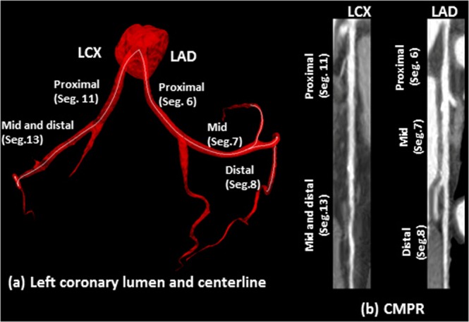
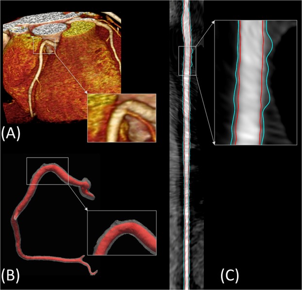
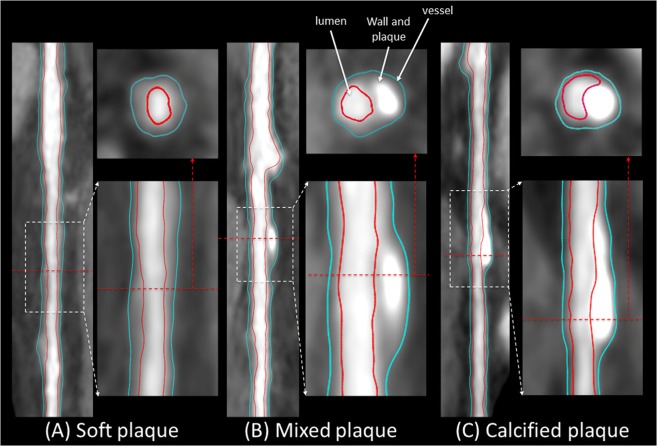
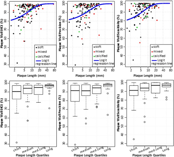
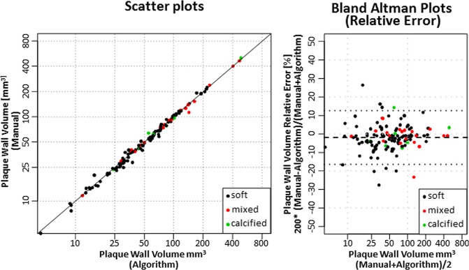
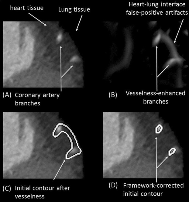
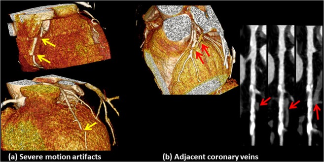
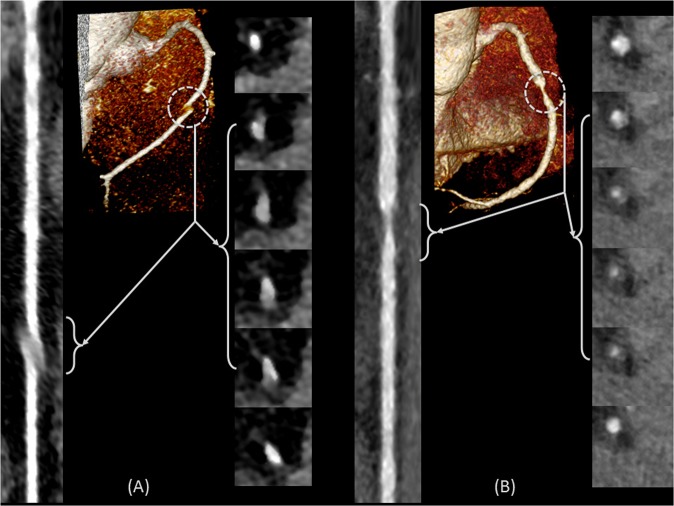
Coronary plaque burden measured by coronary computerized tomography angiography (CCTA), independent of stenosis, is a significant independent predictor of coronary heart disease (CHD) events and mortality. Hence, it is essential to develop comprehensive CCTA plaque quantification beyond existing subjective plaque volume or stenosis scoring methods. The purpose of this study is to develop a framework for automated 3D segmentation of CCTA vessel wall and quantification of atherosclerotic plaque, independent of the amount of stenosis, along with overcoming challenges caused by poor contrast, motion artifacts, severe stenosis, and degradation of image quality. Vesselness, region growing, and two sequential level sets are employed for segmenting the inner and outer wall to prevent artifact-defective segmentation. Lumen and vessel boundaries are joined to create the coronary wall. Curved multiplanar reformation is used to straighten the segmented lumen and wall using lumen centerline. In-vivo evaluation included CCTA stenotic and non-stenotic plaques from 41 asymptomatic subjects with 122 plaques of different characteristics against the individual and consensus of expert readers. Results demonstrate that the framework segmentation performed robustly by providing a reliable working platform for accelerated, objective, and reproducible atherosclerotic plaque characterization beyond subjective assessment of stenosis; can be potentially applicable for monitoring response to therapy.


Conflict of interest statement

The authors declare no competing interests.

Figures

Figure 1
Figure 1
Lumen inner vessel boundary and outer vessel boundary segmentation and visualization block diagram.
Figure 2
Figure 2
An example of the centerline of LCX and LAD (a), which is generated following the segmentation of the inner and outer walls. The centerline is used to generate the straightened arteries using CMPR (b).
Figure 3
Figure 3
Visualization of framework segmentation. (A) The input 3D coronary CTA dataset. (B) Segmented lumen (red) and wall (gray). (C) Straightened vessel demonstrating the delineated inner and outer boundaries of the vessel in red and blue, respectively.
Figure 4
Figure 4
Reformatted coronary artery with (A) soft plaque, (B) mixed plaque, and (C) calcified plaque. The framework-segmented lumen inner contours and vessel outer contours are overlaid in red and blue, respectively.
Figure 5
Figure 5
DICE, precision, and sensitivity performance metrics for wall delineation using algorithm compared to expert observer manual segmentation. Results are plotted vs. plaque length in mm (top row), and vs. plaque length quartiles (bottom row).
Figure 6
Figure 6
Scatter plots and Bland-Altman graphs for the coronary plaque volume delineation using the framework vs. expert manual segmentation.
Figure 7
Figure 7
Improvement in initial contours; avoiding false vesselness artifacts. Coronary branches in the original CCTA (A) and artefactual shapes are enhanced with vesselness (B). Level set segmentation based on vesselness mistakenly includes these false structures (C). Proposed framework correctly identifies the vessel from the other objects (D).
Figure 8
Figure 8
Examples of cases in which the framework will fail due to (a) severe motion artifacts, in which parts of the coronary artery are broken and discontinuous (yellow arrows), and (b) coronary veins that are in close proximity to or in contact with the arteries and are misclassified as plaques (red arrows).
Figure 9
Figure 9
Examples of changes in HU due to motion artifacts (A) and severe stenosis (B), which will lead to level set failure and early termination if used without the proposed initial contouring.
Figure 10
Figure 10
Examples of original CCTA data (left) after the application of the vesselness filter (right). Tubular and sharp-curvature structures are enhanced, which includes vessels and vessel-like structures.
Figure 11
Figure 11
Examples of the calculation of the feature image of the same subject used for the lumen and vessel level set segmentation module at a normal segment (A) and at a stenotic segment (B).

References

    1. Mortality GBD. & Causes of Death, C. Global, regional, and national age-sex specific all-cause and cause-specific mortality for 240 causes of death, 1990–2013: a systematic analysis for the Global Burden of Disease Study 2013. Lancet. 2015;385:117–171. doi: 10.1016/S0140-6736(14)61682-2. - DOI - PMC - PubMed
    1. Falk E, Shah PK, Fuster V. Coronary plaque disruption. Circulation. 1995;92:657–671. doi: 10.1161/01.CIR.92.3.657. - DOI - PubMed
    1. Farb A, et al. Coronary plaque erosion without rupture into a lipid core. A frequent cause of coronary thrombosis in sudden coronary death. Circulation. 1996;93:1354–1363. doi: 10.1161/01.CIR.93.7.1354. - DOI - PubMed
    1. Sangiorgi G, et al. Arterial calcification and not lumen stenosis is highly correlated with atherosclerotic plaque burden in humans: a histologic study of 723 coronary artery segments using nondecalcifying methodology. Journal of the American College of Cardiology. 1998;31:126–133. doi: 10.1016/S0735-1097(97)00443-9. - DOI - PubMed
    1. Glagov S, Weisenberg E, Zarins CK, Stankunavicius R, Kolettis GJ. Compensatory enlargement of human atherosclerotic coronary arteries. The New England journal of medicine. 1987;316:1371–1375. doi: 10.1056/NEJM198705283162204. - DOI - PubMed

Publication types