. 2019 Jan 10;9(1):54.
doi: 10.1038/s41598-018-37185-3.

Structure of the SLy1 SAM homodimer reveals a new interface for SAM domain self-association

Affiliations

Structure of the SLy1 SAM homodimer reveals a new interface for SAM domain self-association

Laura Kukuk et al. Sci Rep. .

Abstract

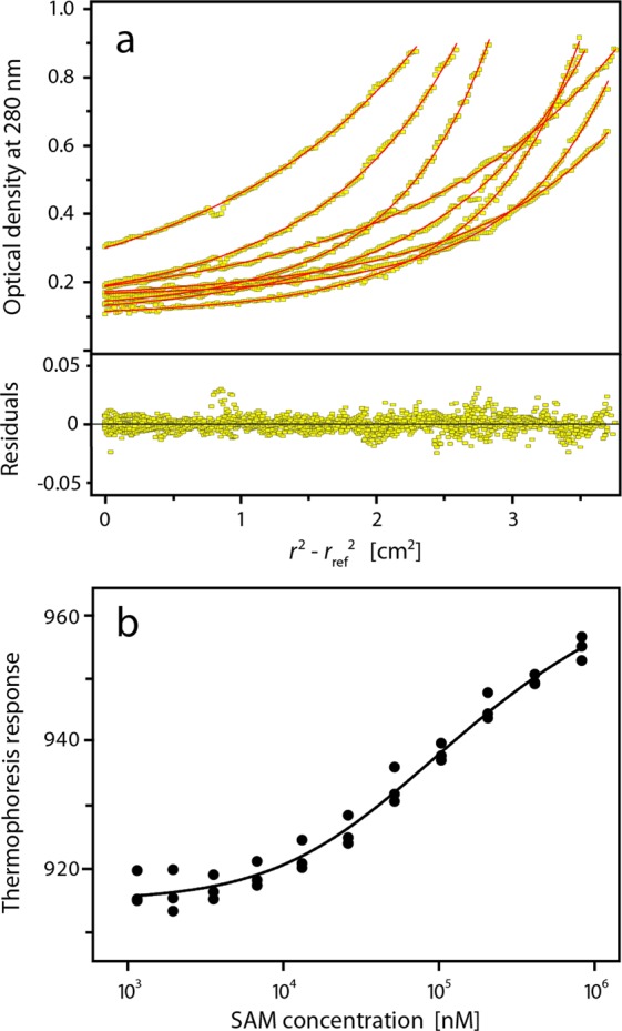
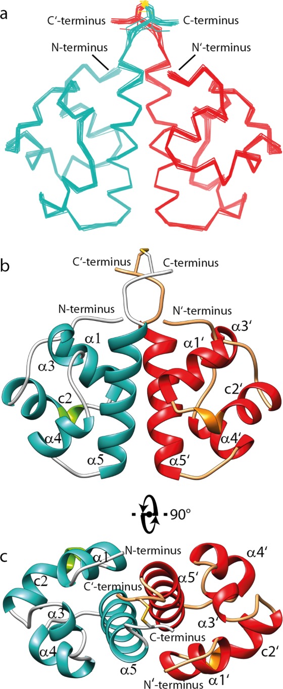
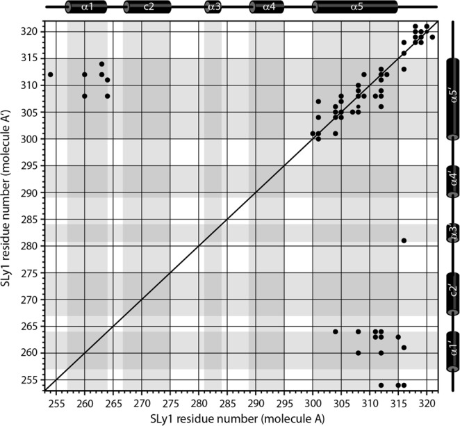
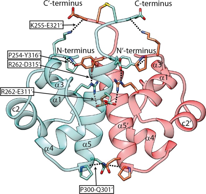
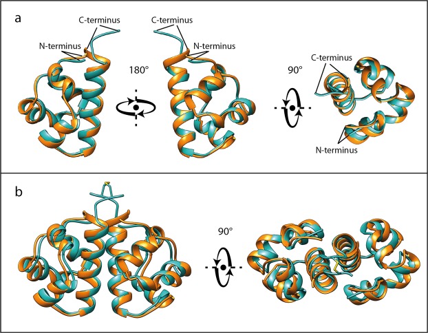
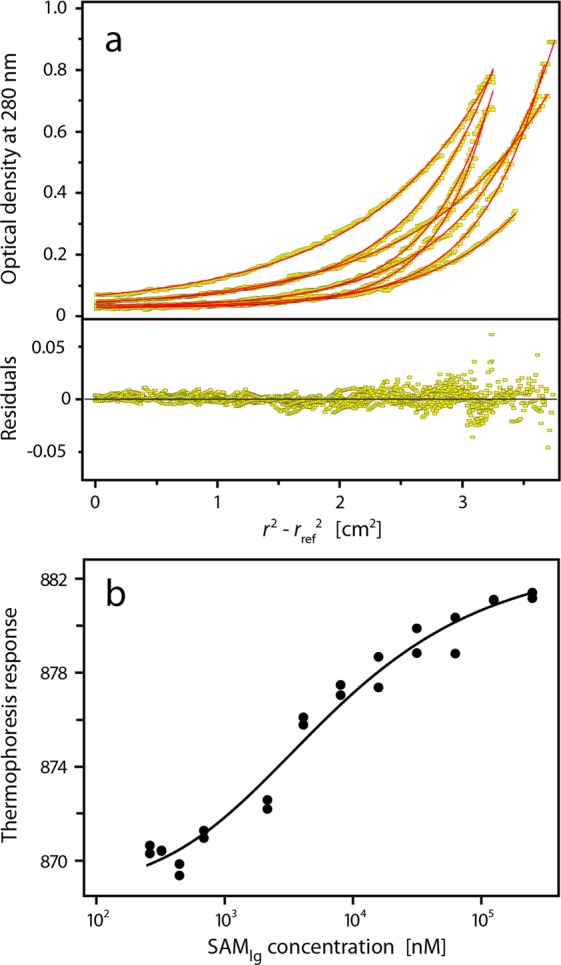
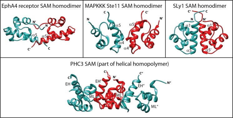
Sterile alpha motif (SAM) domains are protein interaction modules that are involved in a diverse range of biological functions such as transcriptional and translational regulation, cellular signalling, and regulation of developmental processes. SH3 domain-containing protein expressed in lymphocytes 1 (SLy1) is involved in immune regulation and contains a SAM domain of unknown function. In this report, the structure of the SLy1 SAM domain was solved and revealed that this SAM domain forms a symmetric homodimer through a novel interface. The interface consists primarily of the two long C-terminal helices, α5 and α5', of the domains packing against each other. The dimerization is characterized by a dissociation constant in the lower micromolar range. A SLy1 SAM domain construct with an extended N-terminus containing five additional amino acids of the SLy1 sequence further increases the stability of the homodimer, making the SLy1 SAM dimer two orders of magnitude more stable than previously studied SAM homodimers, suggesting that the SLy1 SAM dimerization is of functional significance. The SLy1 SAM homodimer contains an exposed mid-loop surface on each monomer, which may provide a scaffold for mediating interactions with other SAM domain-containing proteins via a typical mid-loop-end-helix interface.


Conflict of interest statement

The authors declare no competing interests.

Figures

Figure 1
Figure 1
Analysis of the SLy1 SAMwt monomer-dimer equilibrium. (a) Sedimentation equilibrium experiments were performed using samples with different SAMwt concentrations (60, 120, and 300 µM) at different speeds. Data were fit globally with a monomer-dimer model. The upper panel shows concentration profiles recorded after establishment of equilibrium between sedimentation and back diffusion and the calculated concentration distributions (red lines) based on a monomer-dimer equilibrium model. The global fit gives a Kd = 117 (33, 423) μM. The lower panel shows the residuals of the fit. (b) Binding isotherm from microscale thermophoresis data. The thermophoresis response of fluorescent labelled SAMwt is dependent on the total concentration of SAMwt. The experimental data were fit to a monomer-dimer equilibrium model (solid line) with a Kd of (153 ± 25) μM.
Figure 2
Figure 2
NMR solution structure of SLy1 SAMC homodimer. (a) Superposition of the 15 lowest energy structures of the disulphide bond-stabilized homodimer of SAMC. The backbones of the monomers are coloured teal and red. (b,c) Ribbon representation of the structure closest to the average backbone structure (r.m.s.d. = 0.19 Å) of the ensemble. α-helices are shown in teal and red in each monomer, whereas the 310-helix part in the composite helix c2 is shown in green and orange. Helices α5 and α5′ are in tight contact to form the major part of the interface. They run in a parallel fashion with an angle of ~−50° between their long axes. Helix α1 and the N-terminus of one monomer are in close proximity to the C-terminal region of helix α5′ of the opposing monomer, and also form part of the dimer interface.
Figure 3
Figure 3
Intermonomer contact pattern in the SLy1 SAMC homodimer. Intermonomer NOE cross correlations (•) in the (13C, 15N) isotope-filtered, 15N- or 13C-edited NOESY spectra from residues in molecule A to residues in molecule A’ Schematic representation of the secondary structure of the SAMC domain is presented above and on the right side of the plot. Shading in the plot defines secondary structure regions.
Figure 4
Figure 4
Surface complementarity of the SAMC monomer. (a) Ribbon and (b) surface representation of the NMR structure of the SAMC monomer are displayed in identical orientation. Surface colouring is based on electrostatic potential at pH 6.4 with negative charges in red and positive charges in blue. Helix α5 forms a hydrophobic ridge with negative charges on the left and hydrophobic residues on the right side. A positively charged ridge formed mainly by side chains of helices α1 and c2 exists. There is a hydrophobic groove between the two ridges. (c) Helix α5′ of the second monomer fits into this hydrophobic groove and side chains E311 and D315 of helix α5′ form salt bridges with residues that are part of the positively charged ridge.
Figure 5
Figure 5
SLy1 SAMC homodimer interface is stabilized by hydrogen bonds and salt bridges. H-bonds and salt bridges between the two monomers of the SAMC homodimer shown in teal and red are labelled. Details on the stabilizing bonds are given in Supplementary Table S9. Side chains of interacting amino acids are shown in stick representation in the ribbon representation of the NMR structure.
Figure 6
Figure 6
Overlay of SAMC NMR and SAMwt X-ray structures. The SAMC structure closest to the average backbone structure of the ensemble of NMR structures of SAMC (teal) and the X-ray structure of SAMwt (orange) are depicted. (a) Superposition of SLy1 SAM monomers (Cα r.m.s.d. = 0.84 Å) is shown. (b) The superposition of the SAMC homodimer with the crystallographic dimer of SAMwt shows nearly identical structures with a Cα r.m.s.d. of 1.13 Å for the dimer.
Figure 7
Figure 7
Analysis of the monomer-dimer equilibrium of SAMlg. (a) Sedimentation equilibrium experiments were performed using samples with different SAMlg concentrations (31, 64, and 98 µM) at multiple speeds. Data were fit globally with a monomer-dimer model. The upper panel shows concentration profiles recorded after establishment of equilibrium between sedimentation and back diffusion and the calculated concentration distributions (red lines) based on a monomer-dimer equilibrium model. The global fit gives a Kd = 2.2 (1.8, 2.6) μM. The lower panel shows the residuals of the fit. (b) Binding isotherm from microscale thermophoresis data. The fit of the experimental data to a monomer-dimer equilibrium model (solid line) yields a Kd of (5.4 ± 1.4) μM.
Figure 8
Figure 8
SAM domain interfaces in homodimers and homopolymers. Identical surfaces of two SAM monomers form the interface of symmetric SAM homodimers. The upper row shows the three types of SAM homodimers that have been reported. The termini-mediated EphA4 receptor SAM homodimer (PDB: 1B0X), the MAPKKK Ste11 SAM homodimer stabilized by interactions between amino acid residues in helices α4 and α5 (PDB: 1X9X), and the SLy1 SAM homodimer stabilized by interactions between amino acid residues in helices α1, α5 and the termini of both monomers (PDB: 6G8O). The monomer-monomer interface in a SAM homopolymer is formed by two different surfaces: the mid-loop (ML) surface of one monomer and the end-helix (EH) surface of the other monomer. This feature enables oligomerization. Three PHC3 SAM monomers that belong to a left-handed helical structure with six monomers per turn (PDB: 4PZO) are displayed in the lower panel.

References

    1. Beer S, Simins AB, Schuster A, Holzmann B. Molecular cloning and characterization of a novel SH3 protein (SLY) preferentially expressed in lymphoid cells. Biochim. Biophys. Acta. 2001;1520:89–93. doi: 10.1016/S0167-4781(01)00242-1. - DOI - PubMed
    1. Astoul E, et al. Approaches to define antigen receptor-induced serine kinase signal transduction pathways. J. Biol. Chem. 2003;278:9267–9275. doi: 10.1074/jbc.M211252200. - DOI - PubMed
    1. Beer S, et al. Impaired immune responses and prolonged allograft survival in Sly1 mutant mice. Molecular and cellular biology. 2005;25:9646–9660. doi: 10.1128/MCB.25.21.9646-9660.2005. - DOI - PMC - PubMed
    1. Reis B, Pfeffer K, Beer-Hammer S. The orphan adapter protein SLY1 as a novel anti-apoptotic protein required for thymocyte development. BMC Immunology. 2009;10:38. doi: 10.1186/1471-2172-10-38. - DOI - PMC - PubMed
    1. Schäll D, Schmitt F, Reis B, Brandt S, Beer-Hammer S. SLy1 regulates T-cell proliferation during Listeria monocytogenes infection in a Foxo1-dependent manner. European J. of Immunol. 2015;45:3087–3097. doi: 10.1002/eji.201545609. - DOI - PubMed

MeSH terms

Substances

LinkOut - more resources