. 2018 Dec 18:9:3126.
doi: 10.3389/fmicb.2018.03126. eCollection 2018.

Venezuelan Equine Encephalitis Virus Capsid Implicated in Infection-Induced Cell Cycle Delay in vitro

Affiliations

Venezuelan Equine Encephalitis Virus Capsid Implicated in Infection-Induced Cell Cycle Delay in vitro

Lindsay Lundberg et al. Front Microbiol. .

Abstract

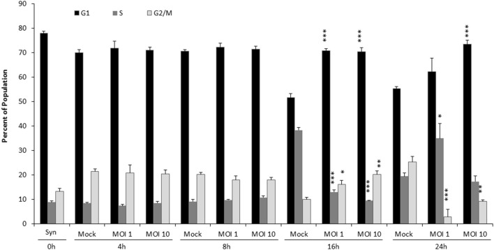
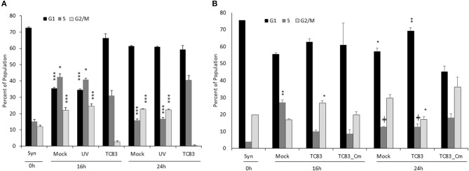
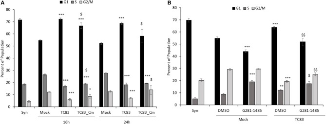
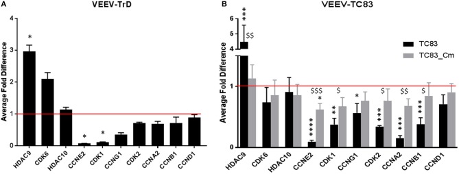
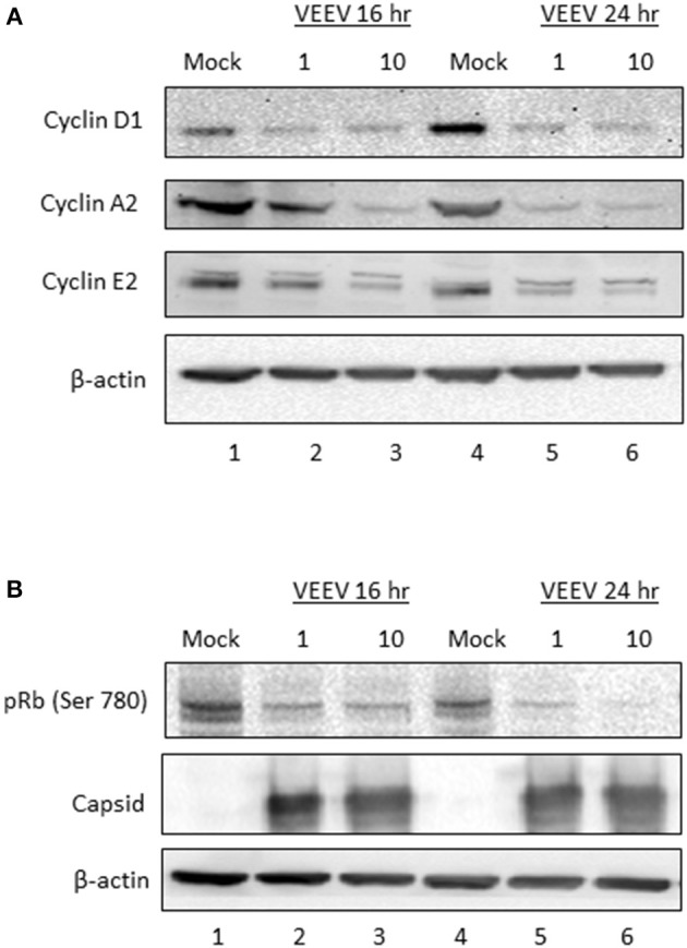
Venezuelan equine encephalitis virus (VEEV) is a positive sense, single-stranded RNA virus and member of the New World alphaviruses. It causes a biphasic febrile illness that can be accompanied by central nervous system involvement and moderate morbidity in humans and severe mortality in equines. The virus has a history of weaponization, lacks FDA-approved therapeutics and vaccines in humans, and is considered a select agent. Like other RNA viruses, VEEV replicates in the cytoplasm of infected cells and eventually induces apoptosis. The capsid protein, which contains a nuclear localization and a nuclear export sequence, induces a shutdown of host transcription and nucleocytoplasmic trafficking. Here we show that infection with VEEV causes a dysregulation of cell cycling and a delay in the G0/G1 phase in Vero cells and U87MG astrocytes. Cells infected with VEEV encoding a capsid NLS mutant or treated with the capsid-importin α interaction inhibitor G281-1485 were partially rescued from this cell cycle dysregulation. Pathway analysis of previously published RNA-sequencing data from VEEV infected U87MG astrocytes identified alterations of canonical pathways involving cell cycle, checkpoint regulation, and proliferation. Multiple cyclins including cyclin D1, cyclin A2 and cyclin E2 and other regulators of the cell cycle were downregulated in infected cells in a capsid NLS dependent manner. Loss of Rb phosphorylation, which is a substrate for cyclin/cdk complexes was also observed. These data demonstrate the importance of capsid nuclear localization and/or importin α binding for inducing cell cycle arrest and transcriptional downregulation of key cell cycle regulators.

Keywords: Venezuelan equine encephalitis virus; alphavirus; capsid; cell cycle; cyclin.


Figures

Figure 1
Figure 1
VEEV TC83 infection delays cell cycle in released, synchronized Vero cells. Vero cells were synchronized via serum-starvation for 72 h. Cells were then infected with TC83 (MOI 1 or 10) or mock-infected for 1 h then released in complete media containing serum. Cells were collected at 4, 8, 16, and 24 hpi, fixed and stained with PI then analyzed for cell cycle by flow cytometry. The average of three biological replicates is displayed. Statistical significance is calculated between mock and infected samples. *p < 0.05, **p < 0.005, ***p < 0.0005.
Figure 2
Figure 2
The cell cycle delay is partially dependent on replicating virus and capsid competent for nuclear import. (A) Vero cells were synchronized via serum-starvation for 72 h. Cells were then infected with TC83 (MOI 1), UV-inactivated TC83, or mock-infected for 1 h then released in complete media containing serum. Cells were collected at 16 and 24 hpi, fixed and stained with PI then analyzed for cell cycle by flow cytometry. The average of three biological replicates is displayed. (B) Similar to (A), Vero cells were infected (MOI 10) with wild-type TC83, TC83_Cm, or mock infected then analyzed by flow cytometry. The average of three biological replicates is displayed, except for the 0 h samples which is N = 1. *Statistical significance compared to mock-infected samples, +Significance compared to TC83_Cm. +p < 0.05, *p < 0.01, **p < 0.001, ***p < 0.0001.
Figure 3
Figure 3
TC83 induces cell cycle delay in U87MG cells which can be relieved by a capsid-importin-α inhibitor. (A) U87MG cells were synchronized via serum-starvation (0.5% FBS) for 72 h. Cells were then infected (MOI 10) with wild-type TC83, TC83_Cm, or mock-infected for 1 h then released in complete media containing 10% FBS. Cells were collected at 16 and 24 hpi, fixed and stained with PI then analyzed for cell cycle by flow cytometry. The average of three biological replicates is displayed. *Statistical significance compared to mock-infected samples, $Significance compared to TC83 infected cells. *p < 0.05, ***p < 0.001, $p < 0.05 (B) Similar to (A), U87MG cells were serum starved (0.1% FBS), treated with DMSO or G281-1485 (10 μM) for 1 h, infected (MOI 10) with wild-type TC83 or mock infected, and post-treated with DMSO or G281-145 in complete media containing 10% FBS. Cells were collected 24 h post-infection and analyzed by flow cytometry. The average of three biological replicates is displayed. *Statistical significance compared to mock-DMSO samples, $Significance compared to TC83-DMSO cells. **p < 0.01, ***p < 0.001. $p < 0.05, $$p < 0.01.
Figure 4
Figure 4
Pathway analysis graph of G1/S checkpoint. Gene expression changes in the G1/S checkpoint pathway generated with IPA. Gene expression values of U87MG cells infected with VEEV-TrD at an MOI 5 at 16 hpi (N = 3 biological replicates) as compared to mock infected cells. Red indicates upregulation and green downregulation of the given gene, and color intensity corresponds to relative difference in gene transcription compared to mock controls. Gray indicates genes that are in the dataset, but did not meet the fold-change or p-value threshold.
Figure 5
Figure 5
RT-qPCR confirmation of RNAseq cell cycle genes in VEEV infected cells. (A) U87MG cells were infected (MOI 5) with VEEV-TrD and then lysed at 16 hpi. After RNA extraction and conversion to cDNA, RT-qPCR was performed for the indicated genes and the fold difference compared to mock infected cells. The average of three biological replicates is displayed. (B) U87MG cells were infected (MOI 10) with TC83 or TC83_Cm then lysed at 16 hpi. After RNA extraction, RT-qPCR was performed for the indicated genes and the fold difference compared to mock infected cells. The average of three biological replicates is displayed. *statistically significant differences from mock, $statistically significant differences from TC83. *p < 0.05, **p < 0.01, ***p < 0.005, $p < 0.05, $$p < 0.01, $$$p < 0.001.
Figure 6
Figure 6
Cyclin protein expression and Rb phosphorylation are decreased following VEEV infection. (A) U87MG cells were synchronized via serum-starvation for 72 h. Cells were then infected (MOI 10) with VEEV TC83 or mock-infected for 1 h and then released in complete media containing 10% FBS. Cells were collected at 16 and 24 hpi for western blot analysis of cyclin D1, E2, A1 or actin expression. (B) Western blot analysis for phospho-Rb, capsid and actin levels in VEEV TC83 or mock infected U87MG cells.
Figure 7
Figure 7
Model of VEEV infection induced transcriptional dysregulation leading to cell cycle delay. Upon VEEV infection, capsid blocks nucleocytoplasmic trafficking through forming a tetrameric complex with CRM1, importin α and importin β. This block results in transcriptional suppression, including an overall decrease in cell cycling associated transcripts, inducing a delay primarily at G0/G1. Nucleocytoplasmic trafficking is intact in uninfected cells. Proteins affected by the block in nucleocytoplasmic trafficking were not determined in this study but are hypothesized to be transcription factors, cell cycle regulators and/or mitotic assembly factors. The specific cyclin and cdk proteins affected following VEEV infection are indicated. Bolded cyclins (cyclin E and A) were decreased at both the mRNA and protein level, whereas expression of the unbolded cyclins and cdks were only assessed at the mRNA level. *Cyclin D mRNA expression was not altered but protein levels were decreased.

References

    1. Atasheva S., Fish A., Fornerod M., Frolova E. I. (2010a). Venezuelan equine encephalitis virus capsid protein forms a tetrameric complex with CRM1 and importin α/β that obstructs nuclear pore complex function. J. Virol. 84, 4158–4171. 10.1128/JVI.02554-09 - DOI - PMC - PubMed
    1. Atasheva S., Garmashova N., Frolov I., Frolova E. (2008). Venezuelan equine encephalitis virus capsid protein inhibits nuclear import in mammalian but not in mosquito cells. J. Virol. 82, 4028–4041. 10.1128/JVI.02330-07 - DOI - PMC - PubMed
    1. Atasheva S., Kim D. Y., Frolova E. I., Frolov I. (2015). Venezuelan equine encephalitis virus variants lacking transcription inhibitory functions demonstrate highly attenuated phenotype. J. Virol. 89, 71–82. 10.1128/JVI.02252-14 - DOI - PMC - PubMed
    1. Atasheva S., Krendelchtchikova V., Liopo A., Frolova E., Frolov I. (2010b). Interplay of acute and persistent infections caused by Venezuelan equine encephalitis virus encoding mutated capsid protein. J. Virol. 84, 10004–10015. 10.1128/JVI.01151-10 - DOI - PMC - PubMed
    1. Baer A., Austin D., Narayanan A., Popova T., Kainulainen M., Bailey C., et al. . (2012). Induction of DNA damage signaling upon Rift Valley fever virus infection results in cell cycle arrest and increased viral replication. J. Bio. Chem. 287, 7399–7410. 10.1074/jbc.M111.296608 - DOI - PMC - PubMed