

Intraductal Papillary Mucinous Neoplasm of the Pancreas Arising in the Setting of an Intermixed Acinar Cell Cystadenoma of the Pancreas: Report of a Rare Case
- PMID: 30631822
- PMCID: PMC6319694
- DOI: 10.1089/crpc.2016.0018
Intraductal Papillary Mucinous Neoplasm of the Pancreas Arising in the Setting of an Intermixed Acinar Cell Cystadenoma of the Pancreas: Report of a Rare Case
Abstract
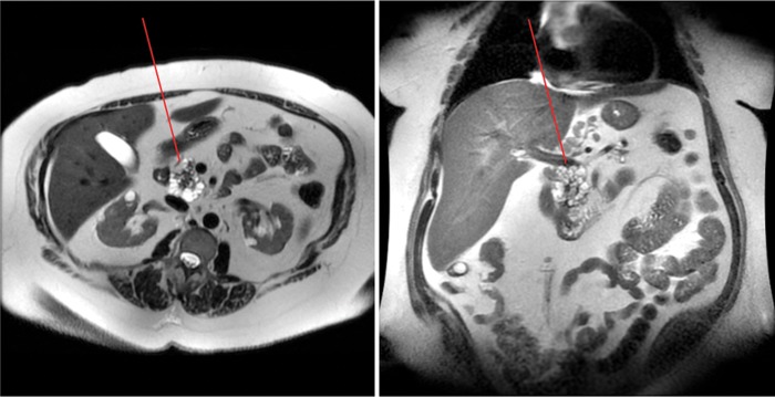
Background: Synchronous cystic lesions of the pancreas with different pathophysiology in the same patient are a rare occurrence.. Case Presentation: We report the incidental finding of a multicystic lesion within the pancreatic head in a morbidly obese woman during workup for bariatric surgery. The lesion contained an intraductal papillary mucinous neoplasm (IPMN) with high-grade dysplasia within an acinar cell cystadenoma (ACA). ACAs are rare tumors first described in 2002. Conclusion: To date, there have been no published reports of synchronous IPMN within an ACA. This case report intends to increase provider awareness of these lesions as well as highlight the importance of surveillance and careful histological examination of heterogeneous cystic lesions of the pancreas.
Keywords: acinar cell cystadenoma; cystic lesions of the pancreas; intraductal papillary mucinous neoplasm; pancreas.
Conflict of interest statement
No competing financial interests exist.
Figures

References
-
- Singhi A, Norwood S, Liu T, et al. . Acinar cell cystadenoma of the pancreas: a benign neoplasm or non-neoplastic ballooning of acinar and ductal epithelium? Am J Surg Pathol 2013;37:1329–1335 - PubMed
-
- Albores-Saavedra J. Acinar cystadenoma of the pancreas: a previously undescribed tumor. Ann Diagn Pathol 2002;6:113–115 - PubMed
-
- Kennedy EP, Brumbaugh J, Yeo CJ. Reconstruction following the pylorus preserving whipple resection: PJ, HJ, and DJ. J Gastrointest Surg 2010;14:408–415 - PubMed
-
- Wolf A, Shirley L, Winter J, et al. . Acinar cell cystadenoma of the pancreas: report of three cases and literature review. J Gastrointest Surg 2013;17:1322–1326 - PubMed
-
- Kim TS, Castillo CF. Diagnosis and management of pancreatic cystic neoplasms. Hematol Oncol Clin North Am 2015;29:655–674 - PubMed
Publication types
LinkOut - more resources
Full Text Sources