Review
. 2019 Mar;266(3):667-679.
doi: 10.1007/s00415-019-09186-3. Epub 2019 Jan 10.

Mills' syndrome revisited

Affiliations
Review

Mills' syndrome revisited

Stephan R Jaiser et al. J Neurol. 2019 Mar.

Abstract

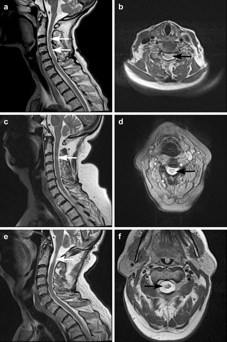
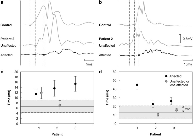
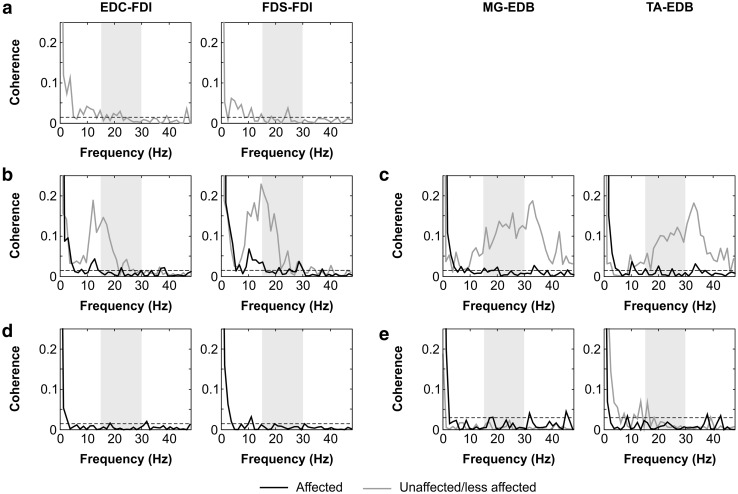
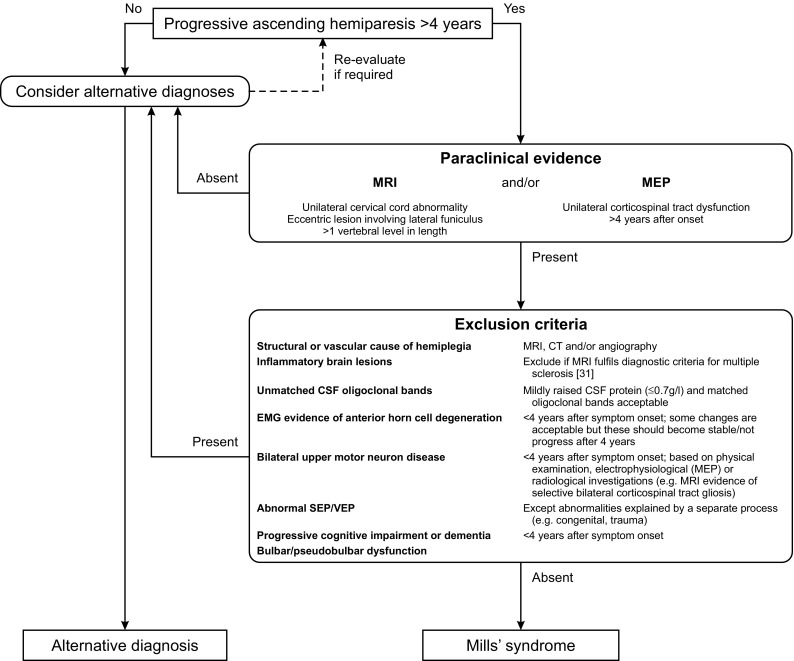
Mills' syndrome is an idiopathic, slowly progressive, spastic hemiparesis. We describe three cases that have been under review for a minimum of 11 years (range 11-19). In all patients, symptoms started in a leg, with a mean age of onset of 59 years (range 53-63). The only abnormality on laboratory investigations was a mildly elevated CSF protein in one case. MRI demonstrated focal T2 hyper-intensity located eccentrically in the cervical cord ipsilateral to the symptomatic side. No cerebral abnormality was demonstrated. Whilst visual and somatosensory evoked potentials were unremarkable, motor evoked potentials were abnormal in all patients: central motor conduction times were significantly prolonged unilaterally in two patients and bilaterally but asymmetrically in the third. Beta-band (15-30 Hz) intermuscular coherence, a potentially more sensitive method of assessing upper motor neuron integrity, was absent unilaterally in one patient and bilaterally in the other two. One patient developed amyotrophy and thus a picture of amyotrophic lateral sclerosis after 16 years, suggesting that Mills' syndrome is part of the motor neuron disease spectrum. Both amyotrophy and subclinical contralateral upper motor neuron disease can therefore be features of Mills' syndrome. However, even with the most sensitive electrodiagnostic techniques, unilateral upper motor neuron disease can remain the only abnormality for as long as 10 years. We conclude that whilst Mills' syndrome should be classified as a motor neuron disorder, it is a distinct nosological entity which can be distinguished from amyotrophic lateral sclerosis, upper motor neuron-dominant amyotrophic lateral sclerosis and primary lateral sclerosis. We propose diagnostic criteria for Mills' syndrome, and estimate a point prevalence of at least 1.2:1,000,000 based on our well-defined referral population in the North of England.

Keywords: MRI; Mills’ syndrome; Neurophysiology; Spastic hemiparesis.


Conflict of interest statement

Conflicts of interest

On behalf of all authors, the corresponding author states that there is no conflict of interest.

Ethical standards

Most investigations were carried out as part of routine clinical care. Studies of motor cortical evoked potentials (MEP) and coherence were approved by the local NHS research ethics committee (approval number 08/H0908/3) and conformed to the Declaration of Helsinki.

Figures

Fig. 1
Fig. 1
MRI findings: sagittal and axial T2 weighted images of the three patients. a, b Patient (1) T2 hyper-intensity in the left lateral aspect of the cervical cord at C3–C5 vertebral levels. c, d Patient (2) focal T2 hyper-intensity involving the left lateral aspect of the cord at C3/4 level. e, f Patient (3) T2 hyper-intensity involving the right anterolateral aspect of the cord at C1/2 level. All three cases have signal abnormality involving the cervical cord which is eccentric in location and involves more than one vertebral level in length
Fig. 2
Fig. 2
Motor evoked potentials (MEP) a, b Examples of average rectified MEP recorded from distal upper limb (first dorsal interosseous) and lower limb (extensor digitorum brevis) muscles. The top traces plotted in grey illustrate MEP from a healthy age-matched control subject. The lower traces are MEP recorded from patient 2 on the clinically affected and unaffected sides. Peripheral motor conduction times were removed and traces then aligned. Apparent latency differences therefore represent differences in central motor conduction times (CMCT). Vertical dashed lines indicate MEP onsets. Note that a shorter timebase has been used in a; the voltage scale bar applies to all MEP shown. c, d Central motor conduction times (CMCT) measured in the upper limb (first dorsal interosseous) and lower limb (extensor digitorum brevis) of each patient. Each data point plots the average of ten individual CMCT measurements taken from unrectified MEP. Error bars represent the standard deviation of the mean. The grey boxes delineate the range within two standard deviations of the mean (solid line) derived from normative CMCT data [9]
Fig. 3
Fig. 3
Intermuscular (EMG–EMG) coherence spectra. Coherence spectra from patients 1 (a), 2 (b, c) and 3 (d, e). Muscle pairs are indicated above each column (extensor digitorum communis (EDC), first dorsal interosseous (FDI), flexor digitorum superficialis (FDS), medial gastrocnemius (MG), extensor digitorum brevis (EDB), and tibialis anterior (TA)). Measurements from the affected limbs are plotted in black, whilst those from the unaffected limbs are plotted in grey. The shaded boxes demarcate the beta-band frequency range of 15–30 Hz. Dashed horizontal lines indicate the 95% significance level. In affected limbs, beta-band coherence was non-significant or borderline in all patients (patient 1 was too weak to perform the upper or lower limb tasks on the affected side). In unaffected limbs, beta-band coherence was retained in patient 2 but absent in patients 1 and 3
Fig. 4
Fig. 4
Proposed diagnostic algorithm for Mills’ syndrome A diagnosis of Mills’ syndrome requires a progressive ascending hemiparesis for at least 4 years, paraclinical evidence for a unilateral corticospinal tract lesion based on MRI or motor evoked potentials (MEP), and the absence of a range of exclusion criteria

References

    1. Armon C, Nelson LM. Is head trauma a risk factor for amyotrophic lateral sclerosis? An evidence based review. Amyotroph Lateral Scler. 2012;13:351–356. doi: 10.3109/17482968.2012.660954. - DOI - PubMed
    1. Bakshi R. Magnetic resonance imaging advances in multiple sclerosis. J Neuroimaging. 2005;15:5S–9S. doi: 10.1177/1051228405283362. - DOI - PubMed
    1. Bäumer D, Butterworth R, Menke RA, Talbot K, Hofer M, Turner MR. Progressive hemiparesis (Mills syndrome) with aphasia in amyotrophic lateral sclerosis. Neurology. 2014;82:457–458. doi: 10.1212/WNL.0000000000000090. - DOI - PMC - PubMed
    1. Bot JCJ, Barkhof F, Polman CH, Lycklama À, Nijeholt GJ, De Groot V, Bergers E, Ader HJ, Castelijns JA. Spinal cord abnormalities in recently diagnosed MS patients: added value of spinal MRI examination. Neurology. 2004;62:226–233. doi: 10.1212/WNL.62.2.226. - DOI - PubMed
    1. Cruz DC, Nelson LM, McGuire V, Longstreth WT., Jr Physical trauma and family history of neurodegenerative diseases in amyotrophic lateral sclerosis: a population-based case-control study. Neuroepidemiology. 1999;18:101–110. doi: 10.1159/000069413. - DOI - PubMed

LinkOut - more resources