. 2019 Feb;15(6):e1804111.
doi: 10.1002/smll.201804111. Epub 2019 Jan 11.

Ultrathin Dual-Scale Nano- and Microporous Membranes for Vascular Transmigration Models

Affiliations

Ultrathin Dual-Scale Nano- and Microporous Membranes for Vascular Transmigration Models

Alec T Salminen et al. Small. 2019 Feb.

Abstract

Selective cellular transmigration across the microvascular endothelium regulates innate and adaptive immune responses, stem cell localization, and cancer cell metastasis. Integration of traditional microporous membranes into microfluidic vascular models permits the rapid assay of transmigration events but suffers from poor reproduction of the cell permeable basement membrane. Current microporous membranes in these systems have large nonporous regions between micropores that inhibit cell communication and nutrient exchange on the basolateral surface reducing their physiological relevance. Here, the use of 100 nm thick continuously nanoporous silicon nitride membranes as a base substrate for lithographic fabrication of 3 µm pores is presented, resulting in a highly porous (≈30%), dual-scale nano- and microporous membrane for use in an improved vascular transmigration model. Ultrathin membranes are patterned using a precision laser writer for cost-effective, rapid micropore design iterations. The optically transparent dual-scale membranes enable complete observation of leukocyte egress across a variety of pore densities. A maximal density of ≈14 micropores per cell is discovered beyond which cell-substrate interactions are compromised giving rise to endothelial cell losses under flow. Addition of a subluminal extracellular matrix rescues cell adhesion, allowing for the creation of shear-primed endothelial barrier models on nearly 30% continuously porous substrates.

Keywords: dual-scale; nanoporous silicon nitride; shear stress; transmigration; vascular mimetic.


Conflict of interest statement

Conflict of Interest

The authors declare the following competing financial interest(s): JLM and TRG are co-founders of SiMPore, an early-stage company commercializing ultrathin silicon-based technologies.

Figures
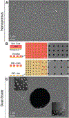
Figure 1.
Figure 1.
Fabrication of dual-scale porous membranes. (A) Scanning electron microscopy (SEM) image of nanoporous silicon nitride membrane prior to the addition of micropores (Scale bar = 1 μm). (B) The addition of micropores begins with the spin coating of Shipley 1805 onto the freestanding nanoporous membrane. The 500 nm coating is developed, forming the micropore pattern (Scale bar = 10 μm). The pattern is transferred into the nanoporous membrane following a reactive ion etch (RIE) process. The RIE ash process removes the Shipley 1805 mask (Scale bar = 10 μm; I,II Scale bars = 4 μm). (C) SEM image of dual-scale (nano- and micro-porous) membrane (Scale bar = 1 μm). Overlaid transmission electron microscopy (TEM) images show further membrane detail including nanopore structure and micropore edge.
Figure 2.
Figure 2.
Micropore etching does not alter nanopores. Transmission electron micrographs show pore structure before (A) and after (B) micropore addition (Scale bars = 50 nm). Histograms of pore size were obtained from lower magnification micrographs to obtain a higher pore distribution. Image analysis was performed in MATLAB to obtain average nanopore diameter, porosity and roundness.
Figure 3.
Figure 3.
Patterning of micropores. (A) Dual-scale membranes were fabricated with eight different micropore patterns and porosities. All micropore patterns are in a square pack configuration. (B) Phase images were recorded of all dual-scale membrane varieties (Scale bar = 20 μm). Specific pore size contributions to total membrane porosity were determined analytically for the highest micropore density (C) and lowest micropore density (D) dual-scale membranes.
Figure 4.
Figure 4.
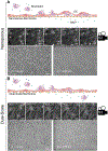
Complete leukocyte transmigration facilitated by dual-scale porous membrane. Time-lapse phase contrast images were recorded of neutrophils migrating through a nanoporous membrane supported human umbilical vein endothelial cell (HUVEC) monolayer (Scale bars = 20 μm). Representative frames are presented. Phase images were taken 2 μm above the membrane (endothelial cell focus) and 430 μm below the membrane (coverslip focus; Scale bar = 200 μm) post-experiment to objectively observe neutrophil complete migration. Experiments were repeated for dual-scale membranes (B). Time-lapse videos can be found in the supplemental material.
Figure 5.
Figure 5.
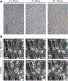
High micropore density leads to endothelial cell loss under fluid shear. HUVECs were seeded on gradient (A) and side-by-side (B) dual-scale (nano- and micro-porous) membranes and grown to confluency. 24 h static images represent microfluidic devices that were replenished with fresh media and returned to the incubator for an additional 24 h. 24 h shear images represent cells that were integrated into the flow circuit and sheared at 4.5 dyn cm−2 for an additional 24 h. Phase and fluorescent (DAPI, blue) images were recorded post-flow (Scale bar = 100 μm) for the same experiment. (C) FIJI assisted quantification of cell nuclei post shear. *Micropore/Cell represents the number of micropores located directly underneath a given endothelial cell based on an estimated spread area of 2000 μm2.
Figure 6.
Figure 6.
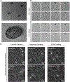
Sub-membrane collagen maintains endothelial monolayer for all micropore densities and permits leukocyte transmigration experimentation. A 300 μm collagen gel was added to the backside of a dual-scale membrane. (A) SEM images were obtained of glutaraldehyde fixed samples without cells (Top Scale bar = 2 μm; Bottom Scale bar = 1 μm). (B) Phase images were obtained of HUVECs seeded on collagen gel back dual-scale membranes and left to grow for 24 h after reaching confluency (Scale bar = 20 μm). (C) Additionally, HUVECs seeded on gel back dual-scale membranes were grown to confluency and sheared at 4.5 dyn cm−2 for 24 h and phase images were recorded (Scale bar = 20 μm). (D) Sub-membrane collagen gel permits neutrophil migration under a fMLP gradient, with an additional ECM crawling step. Roman numerals in panels B and C correspond to the following 3 μm pore densities (pores mm−2): I = 27777, II = 12345, III = 6944, IV = 4444, V = 3086, VI = 2267, VII = 1736, VIII = 1371.
Figure 7.
Figure 7.
Dual-Scale membranes support shear priming for enhanced barrier function in leukocyte migration experimentation. (A) HUVECs were seeded on dual-scale membranes (1736 pores mm−2) and sheared at 4.5 dyn cm−2 for 24 h (Scale bar = 100 μm). (B) Neutrophil migration experiments were performed on the shear primed endothelial cell monolayers. Time-lapse phase contrast imaging reveled some instances of reverse leukocyte migration and probing without complete transendothelial migration.

References

    1. Nourshargh S, Alon R, Immunity 2014, 41 (5), 694–707. - PubMed
    1. Hamilla SM, Stroka KM, Aranda-Espinoza H, PLoS One 2014, 9 (10), e109748. - PMC - PubMed
    1. Teo GSL, Ankrum JA, Martinelli R, Boetto SE, Simms K, Sciuto TE, Dvorak AM, Karp JM, Carman CV, STEM CELLS 2012, 30 (11), 2472–2486. - PMC - PubMed
    1. Teo GSL, Yang Z, Carman CV, Karp JM, Lin CP, STEM CELLS 2014, 33 (1), 265–277. - PMC - PubMed
    1. Weninger W, Biro M, Jain R, Nat Rev Immunol 2014, 14 (4), 232–46. - PubMed

Publication types

LinkOut - more resources