. 2019 Feb 26;13(2):1253-1271.
doi: 10.1021/acsnano.8b06437. Epub 2019 Jan 16.

A Combination of Tri-Leucine and Angiopep-2 Drives a Polyanionic Polymalic Acid Nanodrug Platform Across the Blood-Brain Barrier

A Combination of Tri-Leucine and Angiopep-2 Drives a Polyanionic Polymalic Acid Nanodrug Platform Across the Blood-Brain Barrier

Liron L Israel et al. ACS Nano. .

Abstract

One of the major problems facing the treatment of neurological disorders is the poor delivery of therapeutic agents into the brain. Our goal is to develop a multifunctional and biodegradable nanodrug delivery system that crosses the blood-brain barrier (BBB) to access brain tissues affected by neurological disease. In this study, we synthesized a biodegradable nontoxic β-poly(l-malic acid) (PMLA or P) as a scaffold to chemically bind the BBB crossing peptides Angiopep-2 (AP2), MiniAp-4 (M4), and the transferrin receptor ligands cTfRL and B6. In addition, a trileucine endosome escape unit (LLL) and a fluorescent marker (rhodamine or rh) were attached to the PMLA backbone. The pharmacokinetics, BBB penetration, and biodistribution of nanoconjugates were studied in different brain regions and at multiple time points via optical imaging. The optimal nanoconjugate, P/LLL/AP2/rh, produced significant fluorescence in the parenchyma of cortical layers II/III, the midbrain colliculi, and the hippocampal CA1-3 cellular layers 30 min after a single intravenous injection; clearance was observed after 4 h. The nanoconjugate variant P/LLL/rh lacking AP2, or the variant P/AP2/rh lacking LLL, showed significantly less BBB penetration. The LLL moiety appeared to stabilize the nanoconjugate, while AP2 enhanced BBB penetration. Finally, nanoconjugates containing the peptides M4, cTfRL, and B6 displayed comparably little and/or inconsistent infiltration of brain parenchyma, likely due to reduced trans-BBB movement. P/LLL/AP2/rh can now be functionalized with intra-brain targeting and drug treatment moieties that are aimed at molecular pathways implicated in neurological disorders.

Keywords: Angiopep-2; blood−brain barrier; optical analysis; polymalic acid; targeting peptide; treatment of neurological disorder.


Figures

Figure 1.
Figure 1.
Synthetic route for PMLA/LLL/Angiopep-2/rhodamine (P/LLL/AP2/rh) nanoconjugate production. Biosynthesized PMLA was activated using DCC/NHS chemistry to conjugate LLL and MEA (A). MEA moiety was used to bind AP2 peptide conjugated to a PEG linker (B) via a maleimide-thiol reaction. Rhodamine was attached in the same manner prior to capping with PDP (C).
Figure 2.
Figure 2.
Distinction of nanoconjugate vs lipofuscin fluorescence. Fluorescence in cryosectioned tissue from mice injected with either P/LLL/AP2/rh (A) or PBS (B, C). (A1) Injection of P/LLL/AP2/rh produces diffuse fluorescence in the vascular and perivascular space (asterisks). This fluorescence is only visible if a rhodamine-carrying nanoconjugate is injected and if a rhodamine filter set is used for imaging. (A2) Particulate fluorescence in proximity to DAPI-labeled nuclei is visible in the brain of P/LLL/AP2/rh injected mice (arrows). (B, C) Particulate staining is visible in the brains of PBS injected animals, indicating that particulate fluorescence is related to auto fluorescence (red; lipofuscin). No diffuse perivascular staining is observed in PBS injected mice. (D) Average diameters of lectin-labeled blood vessels for cortical layers II/III (green), the superior/inferior colliculi of the midbrain (red), and the hippocampal CA1-CA3 cellular layers (blue). Data are means and their standard errors from 20 randomly sampled vessels in 4 animals for each brain region. The vessel diameters were measured as the shortest distance between the luminal vessel walls and were 4–5 μm in every brain region.
Figure 3.
Figure 3.
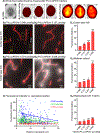
Concentration-dependent BBB penetration of P/LLL/AP2/rh. (A1–A3) Whole-brain epifluorescence images showing absence of fluorescence in PBS injected mice (A1) and incremental fluorescence increases in brains of mice injected with P/LLL/AP2/rh at two different concentrations (as indicated in A2–A4). (B1–B4) Optical imaging of cryosections from brains extracted at 120 min after i.v. injection of P/LLL/AP2/rh at indicated concentrations. The vasculature is shown in red, the nanoconjugate in gray. Drug concentrations are listed with regard to total nanoconjugate content. (C) Nanoconjugate fluorescence intensity vs “distance from vasculature” measurements in brain parenchyma of mice injected with three different concentrations. Fluorescence measurements were obtained from 10 μm2-sized ROI that were randomly overlaid on regions devoid of vasculature (see yellow squares in B1). Intensity measurements and positions were then obtained for each ROI and plotted against the location of the nearest blood vessel wall. (D1–3) Average nanoconjugate fluorescence in the brain parenchyma measured following injections at four different drug concentrations in μmol/kg. Fluorescence is shown as relative intensity, which is the measured nanoconjugate fluorescence intensity after subtraction of autofluorescence intensity measured in PBS injected animals using the same optical imaging and acquisition settings. All statistical tests in D1–3 were conducted against P/LLL/AP2/rh at 0.068 μmol/kg; individual test results are indicated with asterisks where * = p < 0.01, ** = p < 0.001, and *** = p < 0.0001.
Figure 4.
Figure 4.
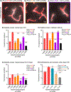
Nanoconjugate composition determines degree and locus of BBB penetration. (A1–3) Optical imaging data showing nanoconjugate permeation of the cerebral cortex: Nanoconjugate fluorescence is gray, and the vasculature is red. Different nanoconjugates are indicated with different colors. (B) Average nanoconjugate fluorescence in layers II/III of the somatosensory cortex (B1), the midbrain colliculi (B2), and the hippocampal CA1–3 cell layers (B3) as a function of nanoconjugate composition and concentration: P/LLL/AP2/rh is shown in red, P/AP2/rh in green, and P/LLL/rh in blue. Average nanoconjugate fluorescence measurements were obtained from 20 randomly sampled ROIs explicitly outside of the cerebral vasculature (4 mice with 4 images each, for each measurement). Statistical tests were conducted between nanoconjugate types (e.g., red vs green) within different concentrations. The results are indicated with asterisks where * = p < 0.01, ** = p < 0.001, and *** = p < 0.0001; the red lines show the concentration of P/LLL/AP2/rh against which each comparison was made.
Figure 5.
Figure 5.
Nanoconjugate peptide moiety screen. P/LLL/rh was equipped with different peptides to assess their role in BBB penetration. (A1–3) Optical imaging data showing rhodamine-labeled nanoconjugate permeation of the cerebral cortex by P/LLL/rh conjugated to AP2 (A1), M4 (A2), and B6 (A3). Nanoconjugate fluorescence is gray and the vasculature is red. Average nanoconjugate fluorescence in layers II/ III of the (B) cerebral cortex, (C) the midbrain colliculi, and (D) hippocampal CA1–3 layers. (E) Nanoconjugate fluorescence measurements in cortical layers II/III for peptide combinations and altered loads of AP2 and rhodamine. Peptide identity is color coded and indicated on the side of the histogram. Statistical tests were conducted against each of the different concentrations of P/LLL/AP2/rh in each histogram and are indicated with asterisks where * = p < 0.01, ** = p < 0.001, and *** = p < 0.0001; the red lines show the concentration of P/LLL/ AP2/rh against which each comparison was made.
Figure 6.
Figure 6.
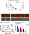
Pharmacokinetics of nanoconjugate fluorescence in serum and brain tissue. (A) Serum clearance analysis was conducted for P/ LLL/AP2/rh (red) and P/LLL/rh (blue) and optically via imaging of the sagittal sinus blood vessel (black). (B) Optical imaging data showing drug clearance and parenchyma accumulation over 240 min. Top images show the nanoconjugate P/LLL/AP2/rh in red and the vasculature in green; the bottom images show only P/LLL/AP2/rh fluorescence. (C) Vascular fluorescence intensity profile for the sagittal sinus vessel as indicated with a yellow line in (B). Time points are color-coded and indicated in the top right corner of the plot. (D) Time dependence of nanoconjugate fluorescence intensity in brain parenchyma for P/LLL/AP2/rh (red), P/LLL/rh (blue), and P/AP2/rh (green) is different from the serum PK kinetics. Fluorescence has a rapid onset and remains quasi-stable for 120 min. Clearance occurs after 240–480 min. All data shown are from the cerebral cortex and are relative fluorescence intensity values that were corrected for background intensities of representative tissues of PBS injected mice.
Figure 7.
Figure 7.
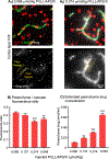
Estimation of the nanoconjugate concentration in nmol/ mL of i.v. injected P/LLL/AP2/rh in the parenchyma of the cerebral cortex. (A1–2) Imaging data showing cortical layer II/III tissue from mice injected with P/LLL/AP2/rh at 0.068 μmol/kg (A1) and 0.274 μmol/kg (A2). The top images show cell nuclei (red), vasculature (green) and P/LLL/AP2/rh (gray). The lower panels show only P/LLL/AP2/rh-associated fluorescence. Yellow and orange ROI indicate how fluorescence was measured in the blood vessels vs the parenchyma: The selected ROIs were close to each other but not ultimately in regions of highest nanoconjugate staining. (B) Fluorescence ratios in vasculature/brain parenchyma. Asterisks indicate statistical significance in Tukey test conducted for the 0.068 μmol/kg drug injection condition, where ** = p < 0.001 and *** = p < 0.0001. (C) Estimated P/LLL/AP2/rh concentration in cortical brain parenchyma. Asterisks indicate statistical significance in Tukey test conducted against the 0.068 μmol/kg drug injection condition, where ** = p < 0.001 and *** = p < 0.0001.
Figure 8.
Figure 8.
BBB penetration of a nanoconjugate carrying D1 peptide. P/LLL/AP2/rh was loaded with a D1 peptide drug load to assess potential effects on BBB penetration. (A1–2) Optical imaging data showing nanoconjugate permeation of the cerebral cortex for P/LLL/ AP2/rh (A1) and P/LLL/AP2/D1/rh (A2). Comparison of the average fluorescence intensity of the two nanoconjugates in layers II/III of the cerebral cortex (B1), the midbrain colliculi (B2), and the hippocampal CA1–3 layers (B3). In each brain region, the intensities are similar for the two conjugates and even significantly increased for P/LLL/AP2/D1/rh in the midbrain colliculi (B2). Statistical results are indicated as *** = p < 0.0001.

References

    1. Dréan A; Goldwirt L; Verreault M; Canney M; Schmitt C; Guehennec J; Delattre J-Y; Carpentier A; Idbaih A Blood-Brain Barrier, Cytotoxic Chemotherapies and Glioblastoma. Expert Rev. Neurother. 2016, 16, 1285–1300. - PubMed
    1. Abbott NJ Blood–Brain Barrier Structure and Function and the Challenges for CNS Drug Delivery. J. Inherited Metab. Dis. 2013, 36, 437–449. - PubMed
    1. Alyautdin R; Khalin I; Nafeeza MI; Haron MH; Kuznetsov D Nanoscale Drug Delivery Systems and the Blood— Brain Barrier. Int. J. Nanomed. 2014, 9, 795–811. - PMC - PubMed
    1. Lopes S; Ribeiro F; Wojnarowicz J; Ƚojkowski W; Jurkschat K; Crossley A; Soares AM; Loureiro S Zinc Oxide Nanoparticles Toxicity to Daphnia Magna: Size-Dependent Effects and Dissolution. Environ. Toxicol. Chem. 2014, 33, 190–198. - PubMed
    1. Shang L; Nienhaus K; Nienhaus GU Engineered Nanoparticles Interacting with Cells: Size Matters. J. Nanobiotechnol. 2014, 12, 5. - PMC - PubMed

Publication types

MeSH terms