

Consequences of the Endogenous N-Glycosylation of Human Ribonuclease 1
- PMID: 30633504
- PMCID: PMC6380942
- DOI: 10.1021/acs.biochem.8b01246
Consequences of the Endogenous N-Glycosylation of Human Ribonuclease 1
Abstract
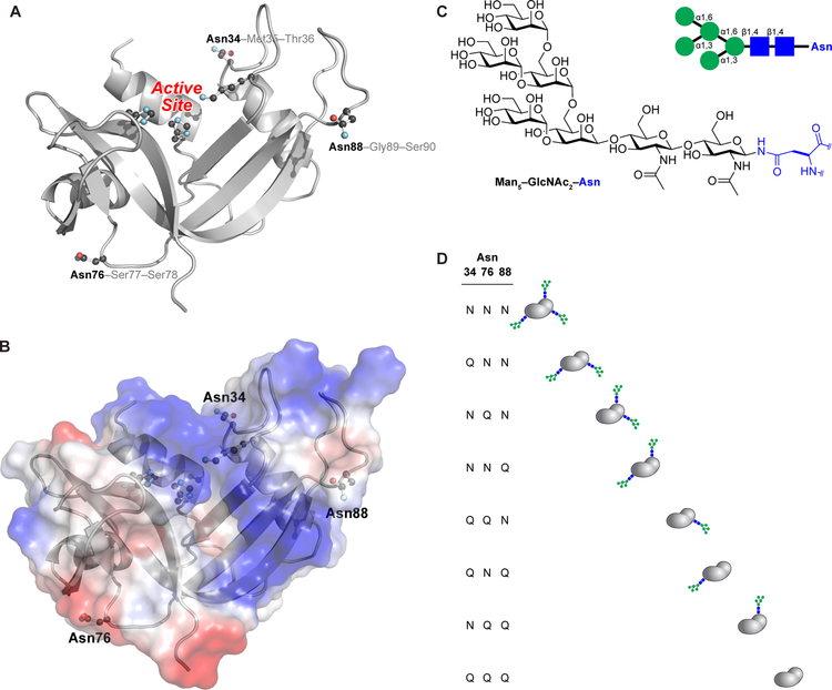
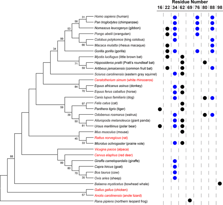
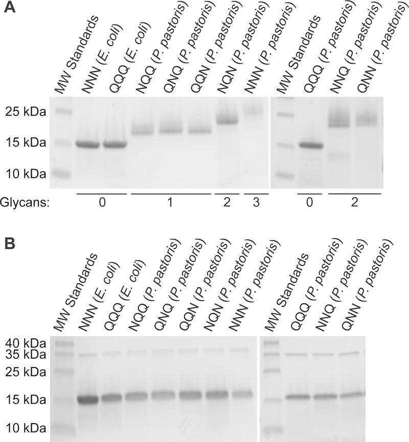
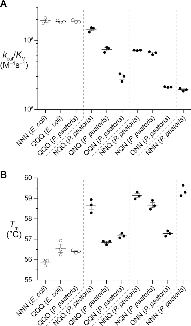
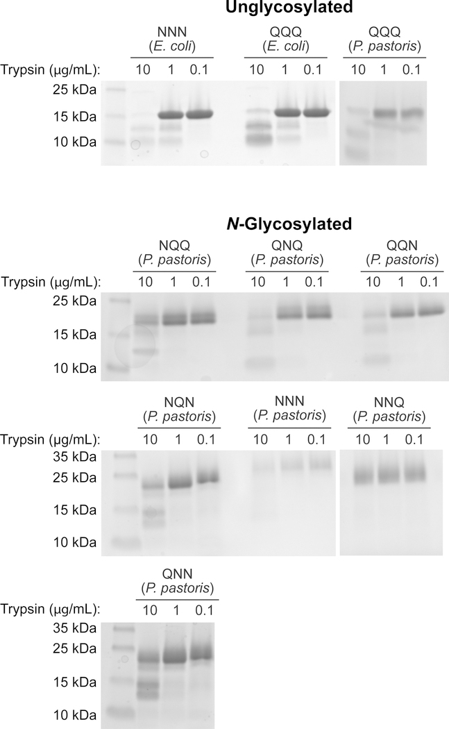
Ribonuclease 1 (RNase 1) is the most prevalent human homologue of the archetypal enzyme RNase A. RNase 1 contains sequons for N-linked glycosylation at Asn34, Asn76, and Asn88 and is N-glycosylated at all three sites in vivo. The effect of N-glycosylation on the structure and function of RNase 1 is unknown. By using an engineered strain of the yeast Pichia pastoris, we installed a heptasaccharide (Man5GlcNAc2) on the side chain of Asn34, Asn76, and Asn88 to produce the authentic triglycosylated form of human RNase 1. As a glutamine residue is not a substrate for cellular oligosaccharyltransferase, we used strategic asparagine-to-glutamine substitutions to produce the three diglycosylated and three monoglycosylated forms of RNase 1. We found that the N-glycosylation of RNase 1 at any position attenuates its catalytic activity but enhances both its thermostability and its resistance to proteolysis. N-Glycosylation at Asn34 generates the most active and stable glycoforms, in accord with its sequon being highly conserved among vertebrate species. These data provide new insight on the biological role of the N-glycosylation of a human secretory enzyme.
Conflict of interest statement
Notes
The authors declare no competing financial interest.
Figures
Similar articles
-
Expression of soluble bovine pancreatic ribonuclease A in Pichia pastoris and its purification and characterization.Biosci Biotechnol Biochem. 2000 Nov;64(11):2437-44. doi: 10.1271/bbb.64.2437. Biosci Biotechnol Biochem. 2000. PMID: 11193413
-
Structure and Dynamics of N-Glycosylated Human Ribonuclease 1.Biochemistry. 2020 Sep 1;59(34):3148-3156. doi: 10.1021/acs.biochem.0c00191. Epub 2020 Jun 30. Biochemistry. 2020. PMID: 32544330 Free PMC article.
-
Degradation of double-stranded RNA by human pancreatic ribonuclease: crucial role of noncatalytic basic amino acid residues.Biochemistry. 2003 Sep 2;42(34):10182-90. doi: 10.1021/bi030040q. Biochemistry. 2003. PMID: 12939146
-
Characterization of N-linked glycosylation on recombinant glycoproteins produced in Pichia pastoris using ESI-MS and MALDI-TOF.Methods Mol Biol. 2009;534:213-23. doi: 10.1007/978-1-59745-022-5_16. Methods Mol Biol. 2009. PMID: 19277549 Review.
-
Emerging biological functions of ribonuclease 1 and angiogenin.Crit Rev Biochem Mol Biol. 2022 Jun;57(3):244-260. doi: 10.1080/10409238.2021.2004577. Epub 2021 Dec 9. Crit Rev Biochem Mol Biol. 2022. PMID: 34886717 Free PMC article. Review.
Cited by
-
Stereoelectronic effects in stabilizing protein-N-glycan interactions revealed by experiment and machine learning.Nat Chem. 2021 May;13(5):480-487. doi: 10.1038/s41557-021-00646-w. Epub 2021 Mar 15. Nat Chem. 2021. PMID: 33723379 Free PMC article.
-
Impact of N-Glycosylation on Protein Structure and Dynamics Linked to Enzymatic C-H Activation in the M. oryzae Lipoxygenase.Biochemistry. 2024 May 21;63(10):1335-1346. doi: 10.1021/acs.biochem.4c00109. Epub 2024 May 1. Biochemistry. 2024. PMID: 38690768 Free PMC article.
-
Human ribonuclease 1 serves as a secretory ligand of ephrin A4 receptor and induces breast tumor initiation.Nat Commun. 2021 May 13;12(1):2788. doi: 10.1038/s41467-021-23075-2. Nat Commun. 2021. PMID: 33986289 Free PMC article.
-
Effects of Lyse-It on endonuclease fragmentation, function and activity.PLoS One. 2019 Sep 30;14(9):e0223008. doi: 10.1371/journal.pone.0223008. eCollection 2019. PLoS One. 2019. PMID: 31568482 Free PMC article.
-
SARS-CoV-2 Spike Protein Post-Translational Modification Landscape and Its Impact on Protein Structure and Function via Computational Prediction.Research (Wash D C). 2023 Mar 8;6:0078. doi: 10.34133/research.0078. Research (Wash D C). 2023. PMID: 36930770 Free PMC article.
References
-
- Sorrentino S (2010) The eight human “canonical” ribonucleases: Molecular diversity, catalytic properties, and special biological actions of the enzyme proteins, FEBS Lett 584, 2194–2200. - PubMed
-
- Kunitz M (1936) Isolation from beef pancreas of a crystalline protein possessing ribonuclease activity, Science 90, 112–113. - PubMed
-
- Richards FM (1972) The 1972 Nobel prize for chemistry, Science 178, 492–493. - PubMed
Publication types
MeSH terms
Substances
Grants and funding
LinkOut - more resources
Full Text Sources
Other Literature Sources
Research Materials
