. 2019 Jan 11;14(1):e0210218.
doi: 10.1371/journal.pone.0210218. eCollection 2019.

Matrix metalloprotease-1 inhibits and disrupts Enterococcus faecalis biofilms

Affiliations

Matrix metalloprotease-1 inhibits and disrupts Enterococcus faecalis biofilms

Lokender Kumar et al. PLoS One. .

Abstract

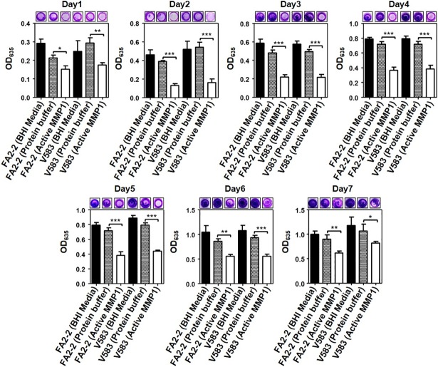
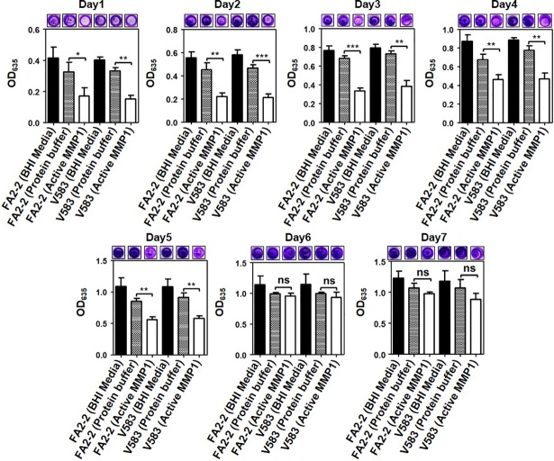
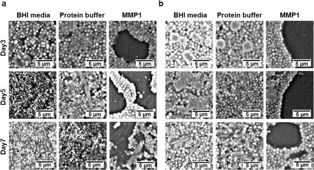
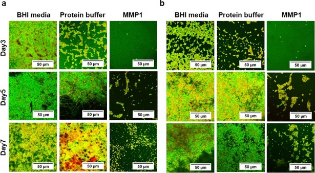
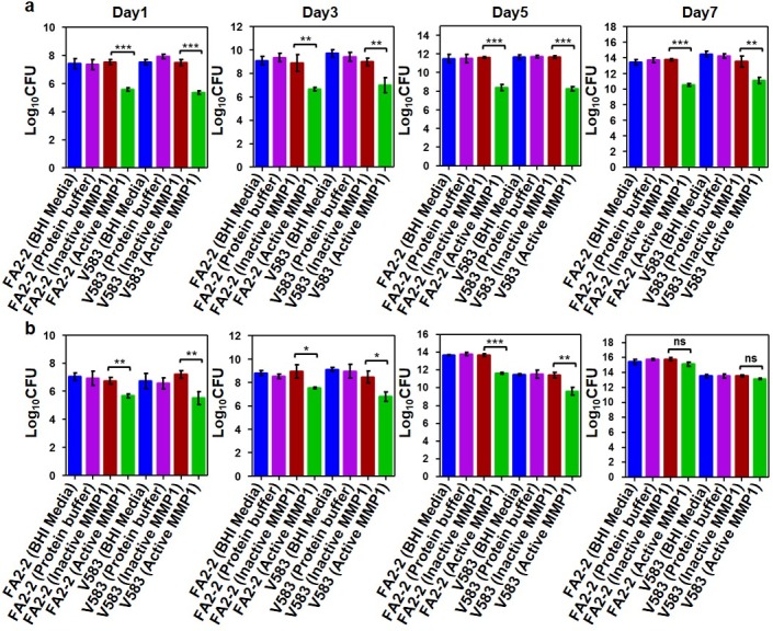
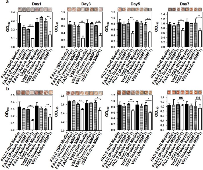
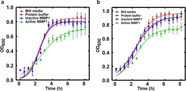
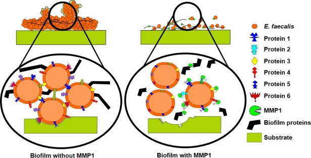
Enterococcus faecalis is a major opportunistic pathogen that readily forms protective biofilms leading to chronic infections. Biofilms protect bacteria from detergent solutions, antimicrobial agents, environmental stress, and effectively make bacteria 10 to 1000-fold more resistant to antibiotic treatment. Extracellular proteins and polysaccharides are primary components of biofilms and play a key role in cell survival, microbial persistence, cellular interaction, and maturation of E. faecalis biofilms. Degradation of biofilm components by mammalian proteases is an effective antibiofilm strategy because proteases are known to degrade bacterial proteins leading to bacterial cell lysis and growth inhibition. Here, we show that human matrix metalloprotease-1 inhibits and disrupts E. faecalis biofilms. MMPs are cell-secreted zinc- and calcium-dependent proteases that degrade and regulate various structural components of the extracellular matrix. Human MMP1 is known to degrade type-1 collagen and can also cleave a wide range of substrates. We found that recombinant human MMP1 significantly inhibited and disrupted biofilms of vancomycin sensitive and vancomycin resistant E. faecalis strains. The mechanism of antibiofilm activity is speculated to be linked with bacterial growth inhibition and degradation of biofilm matrix proteins by MMP1. These findings suggest that human MMP1 can potentially be used as a potent antibiofilm agent against E. faecalis biofilms.


Conflict of interest statement

The authors have declared that no competing interests exist.

Figures

Fig 1
Fig 1. Inhibitory effect of MMP1 on E. faecalis biofilms.
Absorption of CV stain at 635 nm (OD635) of MMP1-treated biofilms of E. faecalis FA2-2 and V583 strains over seven days. Experiments with BHI media and protein buffer were used as controls. For inhibition experiments, biofilms were grown in presence of MMP1 from day0 to day7. Typical images of wells in microtiter plates are given at the top of each panel. * indicates p-value: *<0.01, **<0.01, and ***<0.001. Error bars on the data points represent the standard deviations of 3 technical repeats.
Fig 2
Fig 2. Disruptive effect of MMP1 on E. faecalis biofilms.
Absorption of CV stain at 635 nm (OD635) of MMP1-treated biofilms of FA2-2 and V583 strains over seven days. Experiments with BHI media and protein buffer were used as controls. For disruption experiments, biofilms were first grown in BHI media without MMP1 for desired duration, followed by MMP1 treatment. Typical images of wells in microtiter plates are given at the top of each panel. * indicates p-value: *<0.01, **<0.01, and ***<0.001; ns indicates that the effect is not significant. Error bars on the data points represent the standard deviations of 3 technical repeats.
Fig 3
Fig 3. SEM micrographs of established E. faecalis biofilms.
Biofilms were first grown for 3 to 7 days and then treated with MMP1. (a) Vancomycin susceptible strain FA2-2 and (b) Vancomycin resistant strain V583. In comparison to the control experiments, active MMP1 led to disruption of biofilms resulting in more empty spaces without any bacteria.
Fig 4
Fig 4. Confocal microscopy of E. faecalis biofilms.
Biofilms were grown for 3 to 7 days in the presence of MMP1. (a) Vancomycin susceptible strain FA2-2 and (b) Vancomycin resistant strain V583. Orange and green areas indicate the presence of live and dead bacteria respectively. The amount of live and dead bacteria did not show consistent pattern because of the compounding effects of natural cell death, Triton X100, MMP1, and sample preparation.
Fig 5
Fig 5. Colony forming unit (CFU) assay to quantify viable cells in E. faecalis biofilms.
Quantification (Log 10 CFU) of live bacterial cells in 1, 3 and 7 day old E. faecalis FA2-2 and V583 biofilms treated with and without MMP1 under inhibition (a) and disruption (b) conditions. For inhibition experiments, biofilms were grown in presence of MMP1 from day0 to day7. For disruption experiments, biofilms were first grown in BHI media without MMP1 for desired duration, followed by MMP1 treatment. * indicates p-value: *p<0.01, **p<0.01 and ***p<0.001; ns indicates that the effect is not significant. Error bars on the data points represent the standard deviations of 3 technical repeats.
Fig 6
Fig 6. Congo red staining assay to quantify effects of MMP1 on E. faecalis biofilms.
Absorption of Congo red stain at 490 nm (OD490) of 1, 3 and 7 day old E. faecalis FA2-2 and V583 biofilms treated with and without MMP1 under inhibition (a) and disruption (b) conditions Experiments with BHI media and protein buffer were used as controls. For inhibition experiments, biofilms were grown in presence of MMP1 from day0 to day7. For disruption experiments, biofilms were first grown in BHI media without MMP1 for desired duration, followed by MMP1 treatment. Typical images of wells in microtiter plates are given at the top of each panel. * indicates p-value: *p<0.01, **p<0.01 and ***p<0.001; ns indicates that the effect is not significant. Error bars on the data points represent the standard deviations of 3 technical repeats.
Fig 7
Fig 7. Effect of MMP1 on E. faecalis growth.
Growth curves of E. faecalis in presence of active MMP1, BHI media, and protein buffer for (a) FA2-2 and (b) V583 strains respectively. Symbols represent data points. Solid lines are fits to the logistic equation of bacterial growth, y = a/{1+bexp(−kt)}, where k represents the growth rate. Error bars on the data points represent the standard deviations of 6 technical repeats. For FA2-2 strain, the best fit parameters are: a = 0.85±0.01, b = 24.98±4.44, k = 1.38±0.08 (BHI media); a = 0.81±0.01, b = 32.53±7.02, k = 1.49±0.09 (protein buffer); a = 0.80±0.01, b = 36.62±8.88, k = 1.53±0.10 (inactive MMP1); a = 0.71±0.02, b = 8.47±1.22, k = 0.79±0.06 (active MMP1). For V583 strain, the best fit parameters are: a = 0.96±0.01, b = 13.41±1.20, k = 1.05±0.04 (BHI media); a = 0.92±0.01, b = 7.70±0.62, k = 0.85±0.04 (protein buffer); a = 0.92±0.01, b = 7.73±0.65, k = 0.88±0.04 (inactive MMP1); a = 0.75±0.01, b = 10.12±1.21, k = 0.96±0.05 (MMP1). For fit parameters, error bars represent the standard error of the mean.
Fig 8
Fig 8. Mechanism of MMP1 antibiofilm activity against E. faecalis.
MMP1 degrades cell-associated proteins and biofilm matrix components) leading to poor cell adhesion, bacterial growth inhibition, biofilm inhibition, and disruption (proteins 1–6 are hypothetical biofilm-associated proteins to illustrate the mechanism of MMP1 activity).

References

    1. Control CfD, Prevention. Antibiotic resistance threats in the United States, 2013: Centres for Disease Control and Prevention, US Department of Health and Human Services; 2013.
    1. Munita JM, Arias CA. Mechanisms of Antibiotic Resistance. Microbiology spectrum. 2016;4(2). 10.1128/microbiolspec.VMBF-0016-2015 - DOI - PMC - PubMed
    1. Lebeaux D, Ghigo JM, Beloin C. Biofilm-related infections: bridging the gap between clinical management and fundamental aspects of recalcitrance toward antibiotics. Microbiology and molecular biology reviews: MMBR. 2014;78(3):510–43. 10.1128/MMBR.00013-14 - DOI - PMC - PubMed
    1. Lewis K. Riddle of biofilm resistance. Antimicrobial agents and chemotherapy. 2001;45(4):999–1007. 10.1128/AAC.45.4.999-1007.2001 - DOI - PMC - PubMed
    1. Donlan RM. Biofilm formation: a clinically relevant microbiological process. Clinical infectious diseases: an official publication of the Infectious Diseases Society of America. 2001;33(8):1387–92. 10.1086/322972 . - DOI - PubMed

Publication types

MeSH terms