. 2019 Jan 11;14(1):e0210656.
doi: 10.1371/journal.pone.0210656. eCollection 2019.

Renal chymase-dependent pathway for angiotensin II formation mediated acute kidney injury in a mouse model of aristolochic acid I-induced acute nephropathy

Affiliations

Renal chymase-dependent pathway for angiotensin II formation mediated acute kidney injury in a mouse model of aristolochic acid I-induced acute nephropathy

Wen-Yeh Hsieh et al. PLoS One. .

Abstract

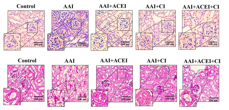
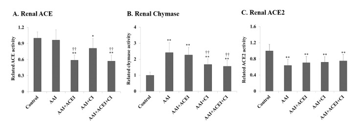
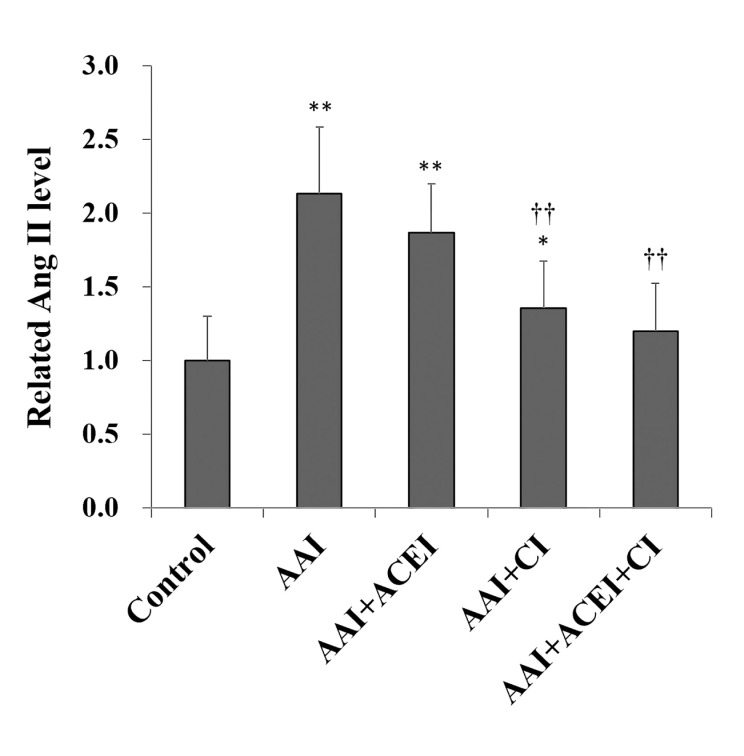
Angiotensin-converting enzyme (ACE) is the primary enzyme that converts angiotensin I (Ang I) to angiotensin II (Ang II) in the renin-angiotensin system (RAS). However, chymase hydrates Ang I to Ang II independently of ACE in some kidney diseases, and it may play an important role. The present study investigated whether chymase played a crucial role in aristolochic acid I (AAI)-induced nephropathy. C57BL/6 mice were treated with AAI via intraperitoneal injection for an accumulated AAI dosage of 45 mg/kg body weight (BW) (15 mg/kg BW per day for 3 days). The animals were sacrificed after acute kidney injury development, and blood, urine and kidneys were harvested for biochemical and molecular assays. Mice exhibited increased serum creatinine, BUN and urinary protein after the AAI challenge. Significant infiltrating inflammatory cells and tubular atrophy were observed in the kidneys, and high immunocytokine levels were detected. Renal RAS-related enzyme activities were measured, and a significantly increased chymase activity and slightly decreased ACE activity were observed in the AAI-treated mice. The renal Ang II level reflected the altered profile of RAS enzymes and was significantly increased in AAI-treated mice. Treatment of AAI-induced nephropathic mice with an ACE inhibitor (ACEI) or chymase inhibitor (CI; chymostatin) reduced renal Ang II levels. The combination of ACEI and CI (ACEI+CI) treatment significantly reversed the AAI-induced changes of Ang II levels and kidney inflammation and injuries. AAI treatment significantly increased renal p-MEK without increasing p-STAT3 and p-Smad3 levels, and p-MEK/p-ERK1/2 signalling pathway was significantly activated. CI and ACEI+CI treatments reduced this AAI-activated signaling pathway. AAI-induced nephropathy progression was significantly mitigated with CI and ACEI+CI treatment. This study elucidates the role of RAS in the pathogenesis of AAI-induced nephropathy.


Conflict of interest statement

The authors have declared that no competing interests exist.

Figures

Fig 1
Fig 1. Body weights, serum and renal biochemical determinations in mice with AAI-induced acute nephropathy.
Animal model of nephropathy was performed in C57BL/6 mice. The mice were treated with AAI via intraperitoneal (i.p.) injection, and the accumulated AAI dosage was 45 mg/kg BW (15 mg/kg BW per day for 3 days). Five groups of 9 mice per group were used. Mouse body weights were recorded daily (A), changes in serum BUN and creatinine (B) levels, and urine protein and urine albumin (C) levels were determined. All values are expressed as the means ± SD of each group (n = 9). ** indicates p < 0.01 compared to the Control group; † and †† indicate p < 0.05 and p < 0.01 compared to the AAI group, respectively.
Fig 2
Fig 2. Renal inflammatory cytokine determinations in mice with AAI-induced acute nephropathy.
Changes in renal IL-6 (A), TGF-β1 (B) and TNF-α (C) levels were determined. All values are expressed as the means ± SD from each group (n = 9). ** indicates p < 0.01 compared to the Control group; † and †† indicate p < 0.05 and p < 0.01 compared to the AAI group, respectively.
Fig 3
Fig 3. Pathological features of AAI-induced kidney injury revealed by light microscopy.
The upper panels show H&E staining and leukocyte infiltration indicative of the inflammation induced by AAI treatment and the alleviation of this response by ACEI, CI and the combination of ACEI and CI (ACEI + CI) treatments. The lower panels show PAS staining and the lesions of tubular atrophy induced by AAI treatment, and the alleviation of lesions by ACEI, CI and ACEI + CI treatments.
Fig 4
Fig 4. Changes in RAS enzyme activity in kidneys of mice with AAI-induced acute nephropathy.
Renal ACE (A), chymase (B) and ACE2 (C) activities were determined, and the related activity levels are shown as levels relative to the Control group. All values are expressed as the means ± SD from each group (n = 9). * and ** indicate p < 0.05 and p < 0.01 compared to the Control group, respectively; †† indicates p < 0.01 compared to the AAI group.
Fig 5
Fig 5. Changes in Angiotensin II (Ang II) in kidneys of mice with AAI-induced acute nephropathy.
Renal Ang II levels were determined, and the related activity levels are shown as the levels relative to the Control group. All values are expressed as the means ± SD from each group (n = 9). * and ** indicate p < 0.05 and p < 0.01 compared to the Control group, respectively; †† indicates p < 0.01 compared to the AAI group.
Fig 6
Fig 6. The level of inflammatory signaling pathways in the kidneys of mice with AAI-induced acute nephropathy.
The expression of p-MEK (A), p-STAT3 (B) and p-Smad3 (C) in kidney tissues was determined using Western blotting and representative results of Western blotting are shown (D). All values are expressed as the means ± SD from each group (n = 9). * and ** indicate p < 0.05 and p < 0.01 compared to the Control group, respectively; †† indicates p < 0.01 compared to the AAI group.
Fig 7
Fig 7. The MAPK signaling pathways in the kidneys of mice with AAI-induced acute nephropathy.
The expression of p-REK1/2 (A), p-JNK (B) and p-38 (C) in kidney tissue was determined using Western blotting, and representative results of Western blotting are shown (D). All values are expressed as the means ± SD from each group (n = 9). * and ** indicate p < 0.05 and p < 0.01 compared to the Control group, respectively; † and †† indicates p < 0.05 and p < 0.01 compared to the AAI group, respectively.

References

    1. Vanherweghem JL, Depierreux M, Tielemans C, Abramowicz D, Dratwa M, Jadoul M, et al. Rapidly progressive interstitial renal fibrosis in young women: association with slimming regimen including Chinese herbs. Lancet. 1993; 341(8842):387–91. - PubMed
    1. Depierreux M, Van Damme B, Vanden Houte K, Vanherweghem JL. Pathologic aspects of a newly described nephropathy related to the prolonged use of Chinese herbs. Am J Kidney Dis. 1994; 24(2):172–80. - PubMed
    1. Jadot I, Declèves AE, Nortier J, Caron N. An integrated view of aristolochic acid nephropathy: update of the literature. Int J Mol Sci. 2017; 18(2):297. - PMC - PubMed
    1. Pailer M, Belohlav L, Simonitsch E. Zur Konstitution der Aristolochiasäuren. Monatsh Chem. 1955; 86(4):676–80.
    1. Solez K, Daugirdas J, Gregory MC, Frohnert PP, Bhowmik DM, Jha V, et al. Is “Chinese herbs nephropathy” a prejudicial term? Am J Kidney Dis. 2001; 38(5):1141–2. - PubMed

Publication types

LinkOut - more resources