Review
. 2020 Jan;51(1):109-138.
doi: 10.1111/ejn.14343. Epub 2019 Jan 29.

Circadian regulation of membrane physiology in neural oscillators throughout the brain

Affiliations
Review

Circadian regulation of membrane physiology in neural oscillators throughout the brain

Jodi R Paul et al. Eur J Neurosci. 2020 Jan.

Abstract

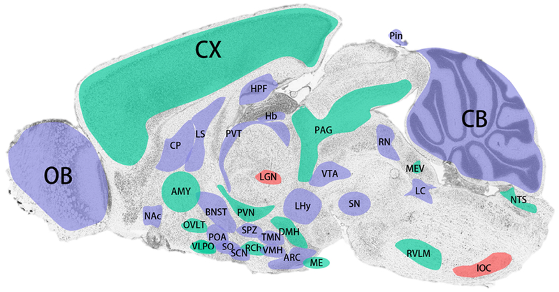
Twenty-four-hour rhythmicity in physiology and behavior are driven by changes in neurophysiological activity that vary across the light-dark and rest-activity cycle. Although this neural code is most prominent in neurons of the primary circadian pacemaker in the suprachiasmatic nucleus (SCN) of the hypothalamus, there are many other regions in the brain where region-specific function and behavioral rhythmicity may be encoded by changes in electrical properties of those neurons. In this review, we explore the existing evidence for molecular clocks and/or neurophysiological rhythms (i.e., 24 hr) in brain regions outside the SCN. In addition, we highlight the brain regions that are ripe for future investigation into the critical role of circadian rhythmicity for local oscillators. For example, the cerebellum expresses rhythmicity in over 2,000 gene transcripts, and yet we know very little about how circadian regulation drives 24-hr changes in the neural coding responsible for motor coordination. Finally, we conclude with a discussion of how our understanding of circadian regulation of electrical properties may yield insight into disease mechanisms which may lead to novel chronotherapeutic strategies in the future.

Keywords: circadian; electrophysiology; extra-SCN; molecular clock; review.


Conflict of interest statement

Conflict of Interest

The authors have no conflicts of interest to disclose.

Image credit: modified from Allen Institute

Figures

Figure 1.
Figure 1.
Illustration of timing molecular rhythms in regions throughout the brain of nocturnal rodents. Where data is available, areas are shaded based on the timing of peak Per1/2 mRNA expression (solid) of WT animals under standard conditions (12:12 LD with ad libitum access to food; variations in light intensity were not taken into account). Areas in which only protein levels (grid pattern) or luciferase assays (striped) have been examined are colored according to the predicted timing of peak Per1/2 expression (i.e. 3 hours before peak protein levels). Gray areas are those that express rhythms but timing is still unknown. Unfilled areas are those which express physiological rhythms, but molecular rhythms are not present or have yet to be explored. The amygdala is colored based on Per2 expression in the central nucleus. For details see text. AMY, amygdala; ARC, arcuate nucleus; BNST, bed nucleus of the stria terminalis; CP, caudate putamen; CX, cortex; DMH, dorsomedial hypothalamus; Hb, habenula; HPF, hippocampal formation; ION, inferior olivary nucleus; LC, locus coeruleus; LGN, lateral geniculate nucleus; LHy, lateral hypothalamus; LS, lateral septum; ME, median eminence; MEV, midbrain trigeminal nucleus; NAc, nucleus accumbens; NTS, nucleus of the solitary tract; OVLT, organum vasculosum of the lamina terminalis; OB, olfactory bulb; PAG, periaqueductal gray; POA, medial preoptic area; PVN, paraventricular nucleus of the hypothalamus; PVT, paraventricular nucleus of the thalamus; RCh, retrochiasmatic area; RN, raphe nuclei; RVLM, rostral ventrolateral medulla; SCN, suprachiasmatic nucleus; SN, substantia nigra; SO, supraoptic nucleus; SPZ, subparaventricular zone; TMN, tuberomammillary nucleus; VMH, ventromedial hypothalamus; VTA, ventral tegmental area.
Figure 2.
Figure 2.
Summary of brain regions expressing molecular and/or physiological rhythms. Areas shaded in red have demonstrated daily rhythms in neuronal excitability. Green shaded areas exhibit oscillations in one or more core molecular clock component. Blue areas are regions which express both molecular and neurophysiological rhythmicity. For details see text. AMY, amygdala; ARC, arcuate nucleus; BNST, bed nucleus of the stria terminalis; CP, caudate putamen; CX, cortex; DMH, dorsomedial hypothalamus; Hb, habenula; HPF, hippocampal formation; IOC, inferior olivary complex; LC, locus coeruleus; LGN, lateral geniculate nucleus; LHy, lateral hypothalamus; LS, lateral septum; ME, median eminence; MEV, midbrain trigeminal nucleus; NAc, nucleus accumbens; NTS, nucleus of the solitary tract; OVLT, organum vasculosum of the lamina terminalis; OB, olfactory bulb; PAG, periaqueductal gray; POA, medial preoptic area; PVN, paraventricular nucleus of the hypothalamus; PVT, paraventricular nucleus of the thalamus; RCh, retrochiasmatic area; RN, raphe nuclei; RVLM, rostral ventrolateral medulla; SCN, suprachiasmatic nucleus; SN, substantia nigra; SO, supraoptic nucleus; SPZ, subparaventricular zone; TMN, tuberomammillary nucleus; VMH, ventromedial hypothalamus; VTA, ventral tegmental area.

References

    1. Abdelsalam S, Uemura H, Umezaki Y, Saifullah AS, Shimohigashi M & Tomioka K (2008) Characterization of PDF-immunoreactive neurons in the optic lobe and cerebral lobe of the cricket, Gryllus bimaculatus. J Insect Physiol, 54, 1205–1212. - PubMed
    1. Abe M, Herzog ED, Yamazaki S, Straume M, Tei H, Sakaki Y, Menaker M & Block GD (2002) Circadian rhythms in isolated brain regions. J Neurosci, 22, 350–356. - PMC - PubMed
    1. Abraham U, Prior JL, Granados-Fuentes D, Piwnica-Worms DR & Herzog ED (2005) Independent circadian oscillations of Period1 in specific brain areas in vivo and in vitro. J Neurosci, 25, 8620–8626. - PMC - PubMed
    1. Adamantidis A & de Lecea L (2009) The hypocretins as sensors for metabolism and arousal. J Physiol, 587, 33–40. - PMC - PubMed
    1. Albers HE, Walton JC, Gamble KL, McNeill J.K.t. & Hummer DL (2017) The dynamics of GABA signaling: Revelations from the circadian pacemaker in the suprachiasmatic nucleus. Front Neuroendocrinol, 44, 35–82. - PMC - PubMed

Publication types