

The application of human amniotic membrane in the surgical management of limbal stem cell deficiency
- PMID: 30633967
- PMCID: PMC6529245
- DOI: 10.1016/j.jtos.2019.01.003
The application of human amniotic membrane in the surgical management of limbal stem cell deficiency
Abstract
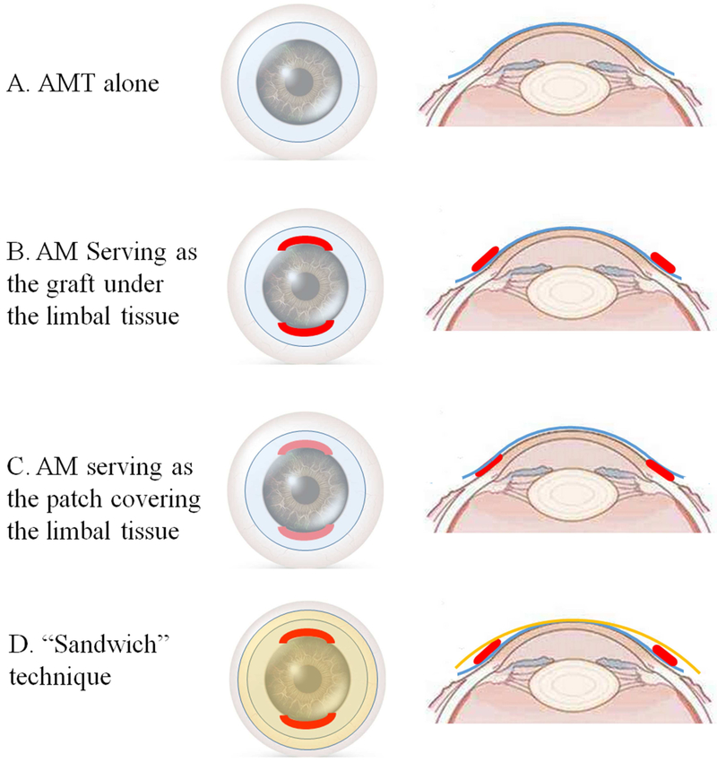
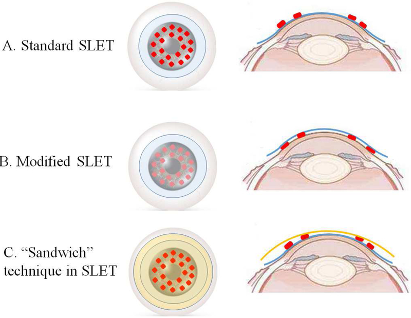
The application of human amniotic membrane (AM) has a wide spectrum of indications in the treatment of ocular surface disorders. Transplantation of AM has been incorporated routinely as a component of ocular surface reconstruction in a variety of ocular pathologies. The application of human AM can be combined with nearly all types of limbal transplantation in treating limbal stem cell deficiency (LSCD). AM provides support and possible protection to the transplanted limbal tissues and limbal stem cells owing to its mechanical and biological properties, and these properties are thought to enhance the success rate of LSC transplantation. This paper reviews the current literature on the applications of AM in the surgical management of LSCD and summarizes the outcome of different surgical approaches. The current literature contains mostly low-level evidences in supporting the role of AM. The efficacy of AM in LSC transplantation needs to be confirmed by randomized controlled clinical trials.
Published by Elsevier Inc.
Figures
References
-
- Fukuda K, Chikama T, Nakamura M, Nishida T. Differential distribution of sub-chains of the basement membrane components type IV collagen and laminin among the amniotic membrane, cornea and conunctiva. Cornea. 1999;18:73–79. - PubMed
-
- Rodriguez-Ares MT, Lopez-Valladares MJ, Tourino R, et al. Effects of lyophilization on human amniotic membrane. Acta Ophthalmol. 2009;87(4):396–403. - PubMed
