. 2019 Mar:136:18-28.
doi: 10.1016/j.ejpb.2019.01.006. Epub 2019 Jan 8.

Polyamidoamine dendrimers-based nanomedicine for combination therapy with siRNA and chemotherapeutics to overcome multidrug resistance

Affiliations

Polyamidoamine dendrimers-based nanomedicine for combination therapy with siRNA and chemotherapeutics to overcome multidrug resistance

Jiayi Pan et al. Eur J Pharm Biopharm. 2019 Mar.

Abstract

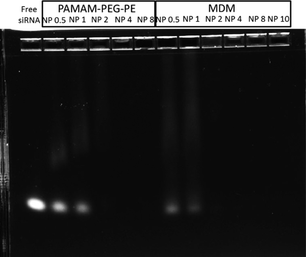
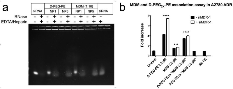
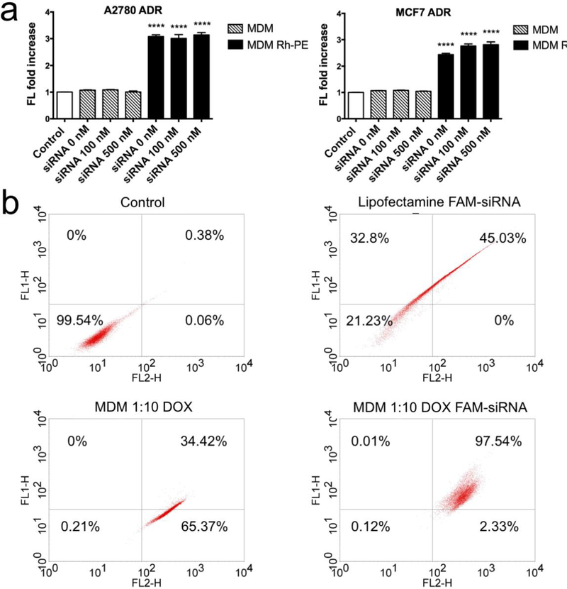
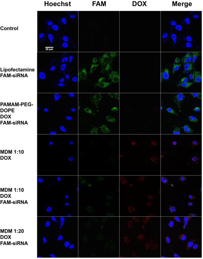
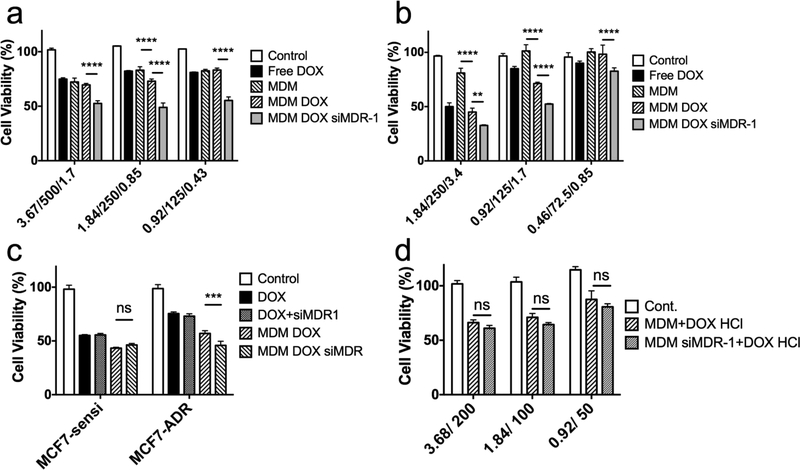
Multidrug resistance (MDR) significantly decreases the therapeutic efficiency of anti-cancer drugs. Its reversal could serve as a potential method to restore the chemotherapeutic efficiency. Downregulation of MDR-related proteins with a small interfering RNA (siRNA) is a promising way to reverse the MDR effect. Additionally, delivery of small molecule therapeutics simultaneously with siRNA can enhance the efficiency of chemotherapy by dual action in MDR cell lines. Here, we conjugated the dendrimer, generation 4 polyamidoamine (G4 PAMAM), with a polyethylene glycol (PEG)-phospholipid copolymer. The amphiphilic conjugates obtained spontaneously self-assembled into a micellar nano-preparation, which can be co-loaded with siRNA onto PAMAM moieties and sparingly water-soluble chemotherapeutics into the lipid hydrophobic core. This system was co-loaded with doxorubicin (DOX) and therapeutic siRNA (siMDR-1) and tested for cytotoxicity against MDR cancer cells: human ovarian carcinoma (A2780 ADR) and breast cancer (MCF7 ADR). The combination nanopreparation effectively downregulated P-gp in MDR cancer cells and reversed the resistance towards DOX.

Keywords: Chemotherapy; Combination delivery; Micelle; Mixed dendrimer micelles (MDM); Multidrug resistance; Polyamidoamine (PAMAM); siRNA delivery.


Conflict of interest statement

Declaration of interest: none

Figures

Figure 1.
Figure 1.
(Colored) Schematic diagram of MDM structure and PAMAM-PEG2k-DOPE.
Figure 2.
Figure 2.
Gel electrophoresis image (2% Agarose gel at 60 mV for 30 mins) indicating the ability of PAMAM-PEG2k-DOPE and MDM 1:20 in complexing siRNA. Full complexations were reached at N/P 2. 750 ng of siRNA was used in each lane.
Figure 3.
Figure 3.
a, Investigation of siRNA protection by PAMAM-PEG2k-DOPE (D-PEG-PE) and MDM using gel electrophoresis. 750 ng of siRNA was used in each lane. b, Cellular association of the conjugate and MDM formulations was checked by flow cytometry using A2780 ADR cell line. Results indicate mean ± SD, n=3. ****p ≤ 0.0001, ***p ≤ 0.001.
Figure 4.
Figure 4.
a. Cellular association assay of MDM 1:10 complexed with 0 nM, 100 nM and 500 nM siMDR-1 in A2780-ADR and MCF7-ADR. MDM Rh-PE are MDMs containing 1% mole of Rh-PE. Results indicate mean ± SD, n=3. ****p ≤ 0.0001. b. Analysis of the co-localization of DOX and FAM-siRNA in MCF7 ADR cell line treated with Lipofectamine FAM-siRNA, MDM 1:10 loaded with DOX and MDM 1:10 co-loaded with DOX and FAM-siRNA using flow cytometry. FL1-H indicates the fluorescence signal at 530 nm. FL2-H indicate the fluorescence signal at 585 nm.
Figure 5.
Figure 5.
(Colored) Confocal images of MCF7 ADR cell line treated with PAMAM-PEG2k-DOPE and different MDM formulations loaded DOX and complexed with FAM-labeled siRNA. Blue: Hoechst 33342; Green: FAM-siRNA; Red: DOX. Magnitude: 63X.
Figure 6.
Figure 6.
a, Characterization of the membrane bound P-gp expression on MCF-7, A2780 cell lines and their Adriamycin resistant counter cell lines using flow cytometry. b, siRNA-mediated P-gp downregulation by MDM 1:10 complexed with siMDR-1 or siRNA control (siNEG) and c, inhibition of P-gp function by MDM 1:10 complexed with siMDR-1 or siRNA Control (siNEG) in the A2780 ADR cell line. Results indicate mean ± SD, n=3. *p ≤ 0.05, **p ≤ 0.01, ***p ≤ 0.001, ****p ≤ 0.0001.
Figure 7.
Figure 7.
Cytotoxicity assay of MDM 1:10 formulations loaded with DOX or DOX/siRNA against a, MCF7 ADR cells and b, A2780 ADR cells. Abscissa labelled “3.67/500/1.7” indicates 3.67 μM nano-preparation loaded with 500 nM siRNA and 1.7 μM DOX. c, Cytotoxicity assay of MCF7 sensitive cell line and MCF7 ADR cell line treated with free DOX 3.4 μM, free DOX 3.4 μM with 200 nM of siMDR-1, MDM 1:10 loaded with 3.4 μM DOX and MDM 1:10 loaded with 3.4 μM DOX and 200 nM siMDR-1. d, Cytotoxicity assay of A2780 ADR cells treated with MDM 1:10 or MDM 1:10 siMDR-1 together with free DOX HCl 3.4 μM. Abscissa labelled “3.68/200” indicates 3.68 μM nano-preparation loaded with 200 nM siRNA. All results indicate mean ± SD, n=3. **P<0.01, ***P<0.001****P<0.0001.

References

    1. Housman G, et al., Drug resistance in cancer: an overview. Cancers (Basel), 2014. 6(3): p. 1769–92. - PMC - PubMed
    1. Sarisozen C, et al., Polymers in the co-delivery of siRNA and anticancer drugs to treat multidrug-resistant tumors. Journal of Pharmaceutical Investigation, 2017. 47(1): p. 37–49.
    1. Rodriguez-Antona C and Ingelman-Sundberg M, Cytochrome P450 pharmacogenetics and cancer. Oncogene, 2006. 25(11): p. 1679–91. - PubMed
    1. Shen H, et al., Comparative metabolic capabilities and inhibitory profiles of CYP2D6.1, CYP2D6.10, and CYP2D6.17. Drug Metab Dispos, 2007. 35(8): p. 1292–300. - PubMed
    1. Townsend DM and Tew KD, The role of glutathione-S-transferase in anti-cancer drug resistance. Oncogene, 2003. 22(47): p. 7369–75. - PMC - PubMed

MeSH terms