Review
. 2019 Jan 9;9(1):17.
doi: 10.3390/biom9010017.

Specialized Roles for Actin in Osteoclasts: Unanswered Questions and Therapeutic Opportunities

Affiliations
Review

Specialized Roles for Actin in Osteoclasts: Unanswered Questions and Therapeutic Opportunities

Guanghong Han et al. Biomolecules. .

Abstract

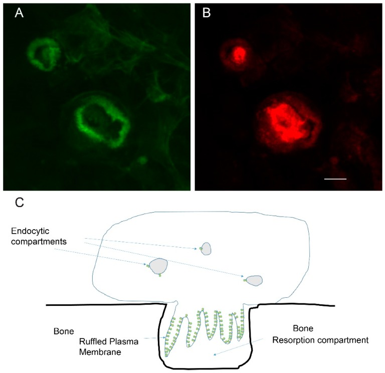
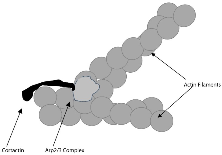
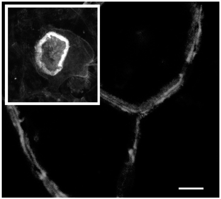
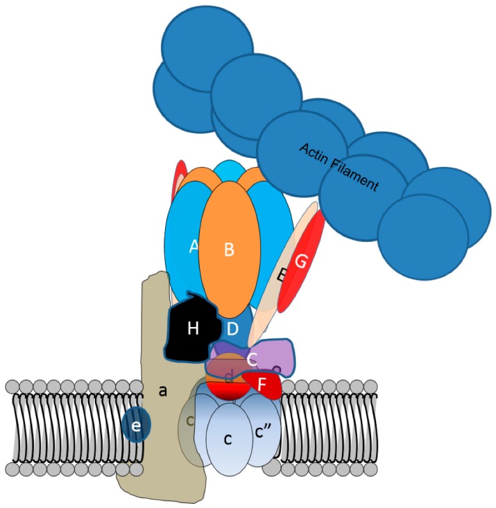
Osteoclasts are cells of the hematopoietic lineage that are specialized to resorb bone. In osteoclasts, the actin cytoskeleton engages in at least two unusual activities that are required for resorption. First, microfilaments form a dynamic and structurally elaborate actin ring. Second, microfilaments bind vacuolar H⁺-ATPase (V-ATPase) and are involved in forming the V-ATPase-rich ruffled plasma membrane. The current review examines these two specialized functions with emphasis on the identification of new therapeutic opportunities. The actin ring is composed of substructures called podosomes that are interwoven to form a cohesive superstructure. Studies examining the regulation of the formation of actin rings and its constituent proteins are reviewed. Areas where there are gaps in the knowledge are highlighted. Microfilaments directly interact with the V-ATPase through an actin binding site in the B2-subunit of V-ATPase. This binding interaction is required for ruffled membrane formation. Recent studies show that an inhibitor of the interaction blocks bone resorption in pre-clinical animal models, including a model of post-menopausal osteoporosis. Because the unusual actin-based resorption complex is unique to osteoclasts and essential for bone resorption, it is likely that deeper understanding of its underlying mechanisms will lead to new approaches to treat bone disease.

Keywords: V-ATPase; actin; actin polymerization; anti-resorptives; bone; bone remodeling; microfilament; vacuolar H+-ATPase.


Conflict of interest statement

The authors declare that they have no conflict of interest.

Figures

Figure 1
Figure 1
Osteoclasts are specialized cells that can resorb mineralized matrix. To do this they form a resorption compartment that depends on generation of an actin ring and ruffled plasma membrane; (A) Confocal micrograph of resorbing osteoclasts on a bone slice stained with phalloidin, which detects filamentous actin, and pseudocolored green. Two actin rings are shown in the field of view; (B) The same cells as in ‘A’ are stained (pseudocolored red) with an antibody that binds the E-subunit of the V-ATPase, a component of the ruffled plasma membrane. The vacuolar H+-ATPase (V-ATPase) is responsible for pumping protons to acidify an extracellular resorption compartment. The scale bar for ‘A’ and ‘B’ is 5 µm; (C) Schematic of a resorbing osteoclast. The ruffled membrane is packed with many V-ATPases (indicated in green). In contrast, endocytic compartments in osteoclasts and other cells require only a few V-ATPases to acidify the compartment. The actin ring, depicted in cross section, is based on actin filaments undergoing rapid polymerization at the cytosolic face of the plasma membrane, and depolymerization toward the center of the cell. The plasma membrane abutted by the actin ring conforms tightly with the bone, creating the extracellular resorption compartment.
Figure 2
Figure 2
A protein complex that includes cortactin and the actin-related protein (Arp) 2/3 complex triggers actin polymerization that forms new filaments at a roughly 70-degree angle from the “mother” filament. This likely forms a base structure of the actin ring.
Figure 3
Figure 3
The actin ring of osteoclast resorbing bone (inset) is thicker, larger, and more dynamic than the actin belt of non-resorbing osteoclasts on a glass coverslip. In the main picture, several large osteoclasts form an almost epithelial like sheet with the actin belt of the contacting cells forming side-by-side. The scale bar is 10 µm for the inset picture and 25 µm for the main picture.
Figure 4
Figure 4
Despite a high affinity actin-binding site in the B-subunit of V-ATPase, normally V-ATPase does not bind microfilaments in cells. This is likely due to the actin binding site being covered by stator arms consisting of the E- and G-subunits. V-ATPase binds microfilaments through an actin binding site in the B2-subunit in osteoclasts when the actin binding site is available. Whether this is due to a change in position of the arm as depicted, or by the loss of one or more stator arms, is not known.
Figure 5
Figure 5
Model suggesting how V-ATPase binding to microfilaments can sort V-ATPase into a vesicle; (A,B) Arp2/3 complex or formin (shown as orange dot) is recruited to a vesicle at a site near V-ATPases and is activated to initiate actin polymerization with actin monomer adding on near the membrane, which is characteristic of actin polymerization in cells; (C) V-ATPase binds tightly to the actin filament at a specific location. As the actin filament elongates, the V-ATPase and associated membrane are drawn away from the vesicle membrane; (D) Eventually, the membrane tether breaks or is actively broken; (E) This leaves a mother vesicle depleted in V-ATPase and a daughter vesicle enriched in V-ATPase. This model is adapted from that of Carnell and colleagues [52]. The same basic mechanism could account for the internalization of V-ATPase from the ruffled plasma membrane.

Similar articles

Cited by

References

    1. Toro E.J., Ostrov D.A., Wronski T.J., Holliday L.S. Rational Identification of Enoxacin as a Novel V-ATPase-Directed Osteoclast Inhibitor. Curr. Protein Pept. Sci. 2012;13:180–191. doi: 10.2174/138920312800493151. - DOI - PMC - PubMed
    1. Teitelbaum S.L. Osteoclasts: what do they do and how do they do it? Am. J. Pathol. 2007;170:427–435. doi: 10.2353/ajpath.2007.060834. - DOI - PMC - PubMed
    1. Blair H.C., Teitelbaum S.L., Ghiselli R., Gluck S. Osteoclastic bone resorption by a polarized vacuolar proton pump. Science. 1989;245:855–857. doi: 10.1126/science.2528207. - DOI - PubMed
    1. Vaananen H.K., Karhukorpi E.K., Sundquist K., Wallmark B., Roininen I., Hentunen T., Tuukkanen J., Lakkakorpi P. Evidence for the presence of a proton pump of the vacuolar H(+)-ATPase type in the ruffled borders of osteoclasts. J. Cell Biol. 1990;111:1305–1311. doi: 10.1083/jcb.111.3.1305. - DOI - PMC - PubMed
    1. Bromme D., Okamoto K., Wang B.B., Biroc S. Human cathepsin O2, a matrix protein-degrading cysteine protease expressed in osteoclasts. Functional expression of human cathepsin O2 in Spodoptera frugiperda and characterization of the enzyme. J. Biol. Chem. 1996;271:2126–2132. doi: 10.1074/jbc.271.4.2126. - DOI - PubMed

Publication types

MeSH terms

Substances

LinkOut - more resources