. 2019 Jan 9;20(2):246.
doi: 10.3390/ijms20020246.

Expression, Cellular and Subcellular Localisation of Kv4.2 and Kv4.3 Channels in the Rodent Hippocampus

Affiliations

Expression, Cellular and Subcellular Localisation of Kv4.2 and Kv4.3 Channels in the Rodent Hippocampus

Rocío Alfaro-Ruíz et al. Int J Mol Sci. .

Abstract

The Kv4 family of voltage-gated K⁺ channels underlie the fast transient (A-type) outward K⁺ current. Although A-type currents are critical to determine somato-dendritic integration in central neurons, relatively little is known about the precise subcellular localisation of the underlying channels in hippocampal circuits. Using histoblot and immunoelectron microscopic techniques, we investigated the expression, regional distribution and subcellular localisation of Kv4.2 and Kv4.3 in the adult brain, as well as the ontogeny of their expression during postnatal development. Histoblot demonstrated that Kv4.2 and Kv4.3 proteins were widely expressed in the brain, with mostly non-overlapping patterns. During development, levels of Kv4.2 and Kv4.3 increased with age but showed marked region- and developmental stage-specific differences. Immunoelectron microscopy showed that labelling for Kv4.2 and Kv4.3 was differentially present in somato-dendritic domains of hippocampal principal cells and interneurons, including the synaptic specialisation. Quantitative analyses indicated that most immunoparticles for Kv4.2 and Kv4.3 were associated with the plasma membrane in dendritic spines and shafts, and that the two channels showed very similar distribution patterns in spines of principal cells and along the surface of granule cells. Our data shed new light on the subcellular localisation of Kv4 channels and provide evidence for their non-uniform distribution over the plasma membrane of hippocampal neurons.

Keywords: electron microscopy; hippocampus; histoblot; immunohistohemistry; potassium channel.


Conflict of interest statement

The authors declare no conflict of interest.

Figures
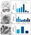
Figure 1
Figure 1
Regional distribution of the Kv4.2 channel in the adult mouse brain. (A,B) The distribution of the Kv4.2 protein was visualised in histoblots of horizontal brain sections at P60 using an affinity-purified anti-Kv4.2 antibody. The expression of Kv4.2 in different brain regions was determined by densitometric analysis of the scanned histoblots. The strongest expression was detected in the cerebellum (Cb) and hippocampus (Hp), with moderate expression in the caudate putamen (CPu) and thalamus (Th). Weak expression level was detected in the cortex (Cx) and septum (Sp); (C,D) In the hippocampus, very strong Kv4.2 expression was detected in the strata oriens (so) and radiatum (sr) of the CA1 region and the molecular layer (ml) of the dentate gyrus (DG); (C,D) Moderate staining was observed in all dendritic layers of CA3, in the stratum lacunosum-moleculare (slm) of the CA1 region and the hilus of the dentate gyrus. Very weak Kv4.2 staining was observed in the stratum pyramidale of the CA1 and CA3 regions and in the granule cell layer of the dentate gyrusso, stratum oriens; sr, stratum radiatum; DG, dentate gyrus; h, hilus.; (E,F) In the cerebellum, the strongest expression level was detected in the granule cell layer (gc), with weak expression in the molecular layer (ml) and very weak in the white matter (wm). Error bars indicate SEM; * p < 0.05; ** p < 0.01; *** p < 0.001. Scale bars: A, 1 mm; C and E, 0.5 mm.
Figure 2
Figure 2
Regional distribution of the Kv4.3 channel in the adult mouse brain. (A,B) The distribution of the Kv4.3 protein was visualised in histoblots of horizontal brain sections at P60 using an affinity-purified anti-Kv4.3 antibody. Kv4.3 expression in different brain regions was determined by densitometric analysis of the scanned histoblots. The strongest expression was detected in the cerebellum (Cb), thalamus (Th) septum (Sp) and hippocampus (Hp), with weaker expression in the cortex (Cx) and caudate putamen (CPu); (C,D) In the hippocampus, strong Kv4.3 expression was detected in the stratum radiatum (sr) of the CA3 region and in the molecular layer (ml) of the dentate gyrus (DG), moderate labelling was observed in all dendritic layers of the CA1 region, the strata oriens (so). Lucidum (sl) and lacunosum-moleculare (slm) of the CA3 region, as well as in the hilus (h) of the dentate gyrus. Very weak Kv4.3 staining was evident in the stratum pyramidale of the CA1 and CA3 regions and in the granule cell layer of the dentate gyrus; (E,F) In the cerebellum, the strongest expression level was detected in the molecular layer (ml), weak expression in the granule cell layer (gc) and very weak in the white matter (wm). Error bars indicate SEM; * p < 0.05; ** p < 0.01. Scale bars: A, 1 mm; C and E, 0.5 mm.
Figure 3
Figure 3
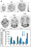
Developmental and regional distribution of the Kv4.2 channel in the mouse brain. (A) Kv4.2 protein distribution was visualised on histoblots of brain horizontal sections at various stages of postnatal development using an affinity-purified anti-Kv4.2 antibody. Kv4.2 was expressed in the brain since the day of birth (P0), and at all stages the strongest expression was detected in the cerebellum (Cb), caudate putamen (CPu), hippocampus (Hp) and thalamus (Th), with the lowest intensity in the cortex (Cx) and septum (Sp); (B) The histoblots were scanned and densitometric measurements from four independent experiments were averaged to compare the protein densities for each developmental time point. The analysis revealed a differential Kv4.2 expression in a developmental stage- and region-specific manner. Kv4.2 expression was detected at P0, increased progressively to reach a peak at P10, P15 or P21 depending on the brain region, and then decreasing at P30. Error bars indicate SEM. Scale bars, 2 mm.
Figure 4
Figure 4
Developmental and regional distribution of the Kv4.3 channel in the mouse brain. (A) Kv4.3 protein distribution was visualised on histoblots of brain horizontal sections at various stages of postnatal development using an affinity-purified anti-Kv4.3 antibody. Kv4.3 was expressed in the brain since the day of birth (P0), and at all stages the strongest expression was detected in the cerebellum (Cb), thalamus (Th), hippocampus (Hp) and septum (Sp), with the lowest intensity in the caudate putamen (CPu) and cortex (Cx); (B) The histoblots were scanned and densitometric measurements from four independent experiments were averaged to compare the protein densities for each developmental time point. The analysis revealed a differential Kv4.3 expression in a region- and developmental-dependent manner. Kv4.3 expression was detected at P0, thought at some time points was not possible to quantify. Then, expression increased progressively to reach a peak at P10, P15 or P21 depending on the brain region, and then decreasing at P30 in cortex, thalamus, caudate putamen and septum, or increasing until P30 in hippocampus and cerebellum. Error bars indicate SEM. Scale bars, 2 mm.
Figure 5
Figure 5
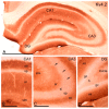
Distribution of immunoreactivity for Kv4.2 in the hippocampus. (A) At the light microscopic level, immunoreactivity for Kv4.2 was widely distributed in the hippocampus but its intensity varied consistently; (B) In the CA1 region, immunolabelling for Kv4.2 was generally moderate-to-strong, with the strata oriens (so) and radiatum (sr) showing the highest and the stratum lacunosum-moleculare (slm) showing weak immunoreactivity. The weakest immunolabelling for Kv4.2 was observed in the stratum pyramidale (sp); (C) In the CA3 region, moderate immunolabelling for Kv4.2 was observed in the strata oriens (so), radiatum (sr) and stratum lacunosum-moleculare (slm), weak in the stratum lucidum and very weak in the stratum pyramidale (sp). Kv4.2 immunolabelling was also seen outlining somata and dendrites of scattered interneurons (arrows); (D) In the dentate gyrus (DG), immunolabelling was strong in the molecular layer (ml), weak in the hilus (h) and the weakest in the granule cell layer (gc). Scale bars: A, 500 µm; BD, 150 µm.
Figure 6
Figure 6
Distribution of immunoreactivity for Kv4.3 in the hippocampus. (A) Immunoreactivity for Kv4.3 was widely distributed in the hippocampus although cellular distribution varied in a subfield-dependent manner; (B) In the CA1 region, immunoreactivity for Kv4.3 was observed exclusively in the soma and dendrites of interneurons distributed throughout the strata oriens (so), pyramidale (sp), radiatum (sr) and lacunosum-moleculare (slm); (C) In the CA3 region, immunolabelling for Kv4.3 was detected along the soma and dendrites of interneurons scattered throughout the strata oriens (so), pyramidale (sp), radiatum (sr) and lacunosum-moleculare (slm). In addition, strong neuropilar staining that indicate expression in pyramidal cells was observed in in strata oriens (so), radiatum (sr) and lacunosum-moleculare (slm) with a weaker labelling in the stratum lucidum (sl), but very weak in the stratum pyramidale (sp); (D) In the dentate gyrus (DG), immunolabelling for Kv4.3 was strong in the neuropil of the molecular layer (mL) and weak in the hilus (h), and also strong in somata and dendrites of scattered interneurons. The granule cell layer of the dentate gyrus showed very weak immunolabelling for Kv4.3. Scale bars: A, 200 µm; BD, 100 µm.
Figure 7
Figure 7
Subcellular localisation of Kv4.2 in the adult hippocampus. Electron micrographs showing immunoparticles for Kv4.2 in the hippocampus, as detected using the pre-embedding immunogold technique at P60. (AD) Immunoreactivity for Kv4.2 was detected in similar neuronal compartments in the CA1 (panels A, B and C) and CA3 (panels D and E) regions and dentate gyrus (DG, panels F and G). Kv4.2 immunoparticles were abundant along the extrasynaptic plasma membrane (arrows) of dendritic spines (s) contacted by axon terminals (at) and dendritic shafts (Den) of pyramidal cells and granule cells. Immunoparticles were also observed at intracellular sites (crossed arrows) in dendritic spines (s) and shafts (Den). Very few immunoparticles for Kv4.2 were also localised to the extrasynaptic plasma membrane (arrowheads) of axon terminals (at) establishing asymmetrical synapses with spines (s). Kv4.2 immunoparticles (double arrowheads) were detected along the main body of the postsynaptic membrane specialisation of GABAergic synapses in the CA1 (panel C), CA3 (panel E) and DG (panel G); (H) Compartmentalisation of Kv4.2 in CA1 pyramidal cells, CA3 pyramidal cells and DG granule cells. Bar graphs showing the percentage of immunoparticles for Kv4.2 at post- and presynaptic compartments, and along the plasma membrane and intracellular sites in dendritic spines, dendritic shafts and axon terminals. A total of 1355 immunoparticles in the CA1, 1223 in the CA3 and 1037 in the DG were analysed. Most immunoparticles were postsynaptic, both along the plasma membrane and at cytoplasmic sites. Postsynaptically, immunoparticles were detected in dendritic spines and in dendritic shafts; (I) Histogram showing the distribution of immunoreactive Kv4.2 in relation to glutamate release sites in dendritic spines of CA1 and CA3 pyramidal cells, and DG granule cells. Data are expressed as the proportion of immunoparticles at a given distance from the edge of the synaptic specialisation. These data show that Kv4.2 immunoparticles were distributed similarly in spines in CA1, CA3 and DG and in the proximity of asymmetrical synapses on dendritic spines. Scale bars: A,D,F,G, 200 nm; B,C,E, 500 nm.
Figure 8
Figure 8
Subcellular localisation of Kv4.3 in the adult hippocampus. Electron micrographs showing immunoparticles for Kv4.3 in the hippocampus, as detected using the pre-embedding immunogold technique at P60. (AC) In the CA1 region, immunoparticles for Kv4.3 were detected along the extrasynaptic plasma membrane (arrows) of dendritic dendritic shafts (Den) of interneurons contacted by both excitatory and inhibitory axon terminals (at) in the strata oriens and radiatum but less frequent in the stratum lacunosum-moleculare. In addition, immunoparticles were observed at cytoplasmic sites (crossed arrows) associated with intracellular membranes. Immunoparticles are present at the edge of the synaptic membrane specialisations (double arrowheads), at synapses between the dendrite (Den) of an interneuron in the stratum oriens and axon terminals (at); (DF) In the CA3 region and DG, immunoreactivity for Kv4.3 was detected along the extrasynaptic plasma membrane (arrows) of interneurons (panel D) or dendritic spines (s) and shafts (Den) of pyramidal cells contacted by axon terminals (at). Few immunoparticles were observed at intracellular sites (crossed arrows) in dendritic spines and shafts, and even fewer (arrowheads) presynaptically in axon terminals (at); (G) Compartmentalisation of Kv4.3 in CA3 pyramidal cells and DG granule cells. Bar graphs showing the percentage of immunoparticles for Kv4.3 at post- and presynaptic compartments, and along the plasma membrane and intracellular sites in dendritic spines, dendritic shafts and axon terminals. A total of 3075 immunoparticles in the CA3 region and 2607 immunoparticles in the DG were analysed. Most immunoparticles were postsynaptic, both along the plasma membrane and at cytoplasmic sites. Postsynaptically, immunoparticles were detected in dendritic spines and in dendritic shafts; (H) Histogram showing the distribution of immunoreactive Kv4.3 in relation to glutamate release sites in dendritic spines of CA3 pyramidal cells and DG granule cells. Data are expressed as the proportion of immunoparticles at a given distance from the edge of the synaptic specialisation. These data show that Kv4.3 immunoparticles were distributed identically in spines of the CA3 and DG and in the proximity of asymmetrical synapses. Scale bars: A,B,D,F, 500 nm; C,E, 200 nm.
Figure 9
Figure 9
Co-localisation of Kv4.2 and Kv4.3 in granule cells. (AC) Electron micrographs showing co-localisation for Kv4.2 with Kv4.3, as detected using a double-labelling pre-embedding immunogold method at P60. Labelling is shown with immunoperoxidase reaction for Kv4.2, and with silver-intensified immunogold reaction for Kv4.3. Immunoparticles for Kv4.3 were seen in dendritic spines (s) and shafts (Den) of granule cells immunopositive for Kv4.2, both along the plasma membrane (arrows) and at intracellular sites (crossed arrows); (D) Change in the density of Kv4.2 and Kv4.3 in DG granule cells as a function of distance from the soma in six somato-dendritic domains. Density of immunoparticles for the two channel subtypes increased significantly from soma to dendritic spines (Sp) in the inner one-third and outer two-thirds of the molecular layer. This analysis demonstrated their similar non-uniform distributions over the neuronal surface of granule cells. Inner SB, spiny branchlets in the inner one-third; Outer SB, spiny branchlets in the outer two-third; Inner Sp, spines in the inner one-third; Outer Sp, spines in the outer two-third. Scale bars: AC, 500 nm.

Similar articles

Cited by

References

    1. Anderson P., Morris R., Amaral D., Bliss T., O’Kefefe J. The Hippocampus Book. Oxford University Press Inc.; New York, NY, USA: 2007.
    1. Poolos N.P., Johnston D. Dendritic ion channelopathy in acquired epilepsy. Epilepsia. 2012;53(Suppl. 9):32–40. doi: 10.1111/epi.12033. - DOI - PMC - PubMed
    1. MacDonald J.F., Belrose J.C., Xie Y.F., Jackson M.F. Nonselective cation channels and links to hippocampal ischemia, aging, and dementia. Adv. Exp. Med. Biol. 2013;961:433–447. - PubMed
    1. Trimmer J.S. Subcellular localization of K+ channels in mammalian brain neurons: Remarkable precision in the midst of extraordinary complexity. Neuron. 2015;85:238–256. doi: 10.1016/j.neuron.2014.12.042. - DOI - PMC - PubMed
    1. Hille B. Ionic Channels of Excitable Membranes. Sinauer Associates Inc.; Sunderland, MA, USA: 2001.

Substances

LinkOut - more resources