. 2019 Jan 10;8(1):68.
doi: 10.3390/jcm8010068.

Muscle Involvement in a Large Cohort of Pediatric Patients with Genetic Diagnosis of Mitochondrial Disease

Affiliations

Muscle Involvement in a Large Cohort of Pediatric Patients with Genetic Diagnosis of Mitochondrial Disease

Cristina Jou et al. J Clin Med. .

Abstract

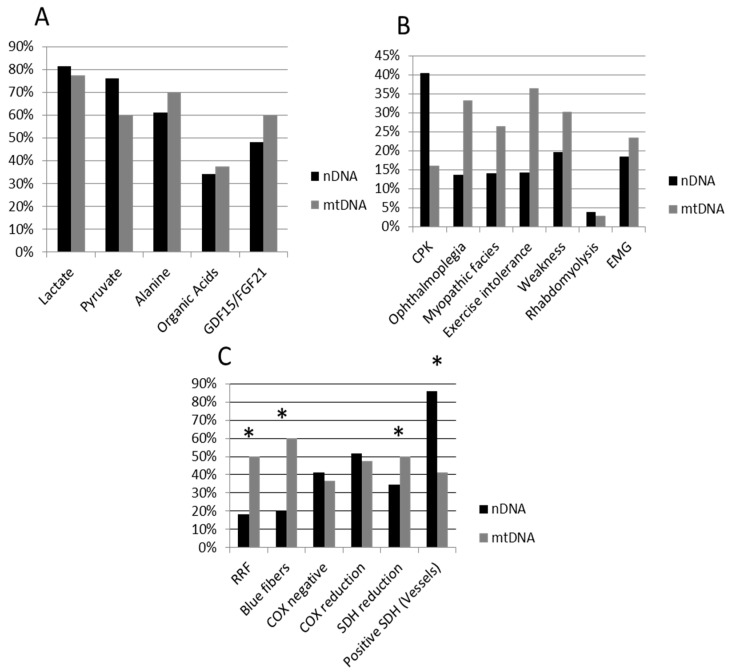
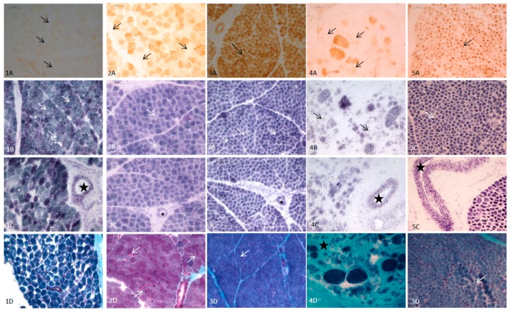
Mitochondrial diseases (MD) are a group of genetic and acquired disorders which present significant diagnostic challenges. Here we report the disease characteristics of a large cohort of pediatric MD patients (n = 95) with a definitive genetic diagnosis, giving special emphasis on clinical muscle involvement, biochemical and histopathological features. Of the whole cohort, 51 patients harbored mutations in nuclear DNA (nDNA) genes and 44 patients had mutations in mitochondrial DNA (mtDNA) genes. The nDNA patients were more likely to have a reduction in muscle fiber succinate dehydrogenase (SDH) stains and in SDH-positive blood vessels, while a higher frequency of mtDNA patients had ragged red (RRF) and blue fibers. The presence of positive histopathological features was associated with ophthalmoplegia, myopathic facies, weakness and exercise intolerance. In 17 patients younger than two years of age, RRF and blue fibers were observed only in one case, six cases presented cytochrome c oxidase (COX) reduction/COX-fibers, SDH reduction was observed in five and all except one presented SDH-positive blood vessels. In conclusion, muscle involvement was a frequent finding in our series of MD patients, especially in those harboring mutations in mtDNA genes.

Keywords: biochemical markers; mitochondrial diseases; muscle histopathology; myopathy; next generation sequencing; pediatric patients.


Conflict of interest statement

The authors declare no conflict of interests.

Figures

Figure 1
Figure 1
Percentage of impaired biomarkers (A), muscular involvement (B) and positive histopathological features (C) in nDNA (black bars) and mtDNA (grey bars) patients. Whiskers indicate significant differences (chi-square test) between nDNA and mtDNA groups. nDNA patients showed significantly higher frequency of SDH reduction and SDH-positive vessels, while mtDNA patients shower a higher frequency of RRF and blue fibers, and near significative for ophthalmoplegia and exercise intolerance. nDNA, nuclear DNA; mtDNA, mitochondrial DNA; CPK, creatine phosphate kinase; EMG, myopathic electromyography; RRF, ragged red; SDH, succinate dehydrogenase; COX, cytochrome c oxidase.
Figure 2
Figure 2
Main histopathological features (quadriceps muscle) of 5 infants with mitochondrial disease (MD) and other congenital myopathies. (1) Patient with AGK mutations (3 days old); (1A): Total absence of COX (arrows). (1B): Normal stain of SDH (arrows). (1C): SDH-positive vessels (star). (1D): Modified Gomori trichrome: No RRF (arrow marks a normal fiber). (2) Patient with SUCLA2 mutations (6 months old); (2A): Isolated COX negative fibers (arrows). (2B): Mild reduction of SDH (arrows). (2C): SDH-positive vessels (star). (2D) Modified Gomori trichrome: occasional subsarcolemal mitochondrial proliferation (arrows). (3) Patient with MT-ATP6 mutations (1 month old); (3A): COX: Muscular fibers with a normal pattern showing the highest intensity of stain in type I fibers, and pale and intermediate intensity in type II fibers (arrow mark a type I muscular fiber). (3B): Muscular fibers with normal SDH stain. (3C): SDH-positive vessels (star). (3D): Muscular fibers with normal pattern and no RRF with modified Gomori trichrome stain. (4) Patient with LAMA2 mutations (6 months old) (4A): Isolated fibers with COX reduction consistent with pre-necrotic fibers (arrows). (4B): Isolated fibers with SDH reduction (arrows). (4C): SDH-positive vessels (star). (4D): Modified Gomori trichrome; marked increase of endomysial tissue (star) consistent with dystrophic pattern, no RRF. (5) Patient with miotubularin mutation (1 month old). (5A): No COX negativefibers (arrows). (5B): No reduction of SDH (arrows). (5C): SDH-positive vessels (star). (5D): Modified Gomori trichrome. Large central nuclei in several fibers (arrows). No RRF.

Similar articles

Cited by

References

    1. Witters P., Saada A., Honzik T., Tesarova M., Kleinle S., Horvath R., Goldstein A., Morava E. Revisiting mitochondrial diagnostic criteria in the new era of genomics. Genet. Med. 2018;20:444–451. doi: 10.1038/gim.2017.125. - DOI - PubMed
    1. Lightowlers R.N., Taylor R.W., Turnbull D.M. Mutations causing mitocondrial disease: What is new and what challenges remain? Science. 2015;349:1494–1499. doi: 10.1126/science.aac7516. - DOI - PubMed
    1. Morava E., van den Heuvel L., Hol F., de Vries M.C., Hogeveen M., Rodenburg R.J., Smeitink J.A. Mitochondrial disease criteria: Diagnostic applications in children. Neurology. 2006;67:1823–1826. doi: 10.1212/01.wnl.0000244435.27645.54. - DOI - PubMed
    1. Phadke R. Myopathology of adult and paediatric mitochondrial diseases. J. Clin. Med. 2017;6:64. doi: 10.3390/jcm6070064. - DOI - PMC - PubMed
    1. Menezes M.J., Riley L.G., Christodoulou J. Mitochondrial respiratory chain disorders in childhood: Insights into diagnosis and management in the new era of genomic medicine. Biochim. Biophys. Acta. 2014;1840:1368–1379. doi: 10.1016/j.bbagen.2013.12.025. - DOI - PubMed

LinkOut - more resources