. 2019 Jan 10;11(1):69.
doi: 10.3390/cancers11010069.

Functional Assessment for Clinical Use of Serum-Free Adapted NK-92 Cells

Affiliations

Functional Assessment for Clinical Use of Serum-Free Adapted NK-92 Cells

Michael Chrobok et al. Cancers (Basel). .

Abstract

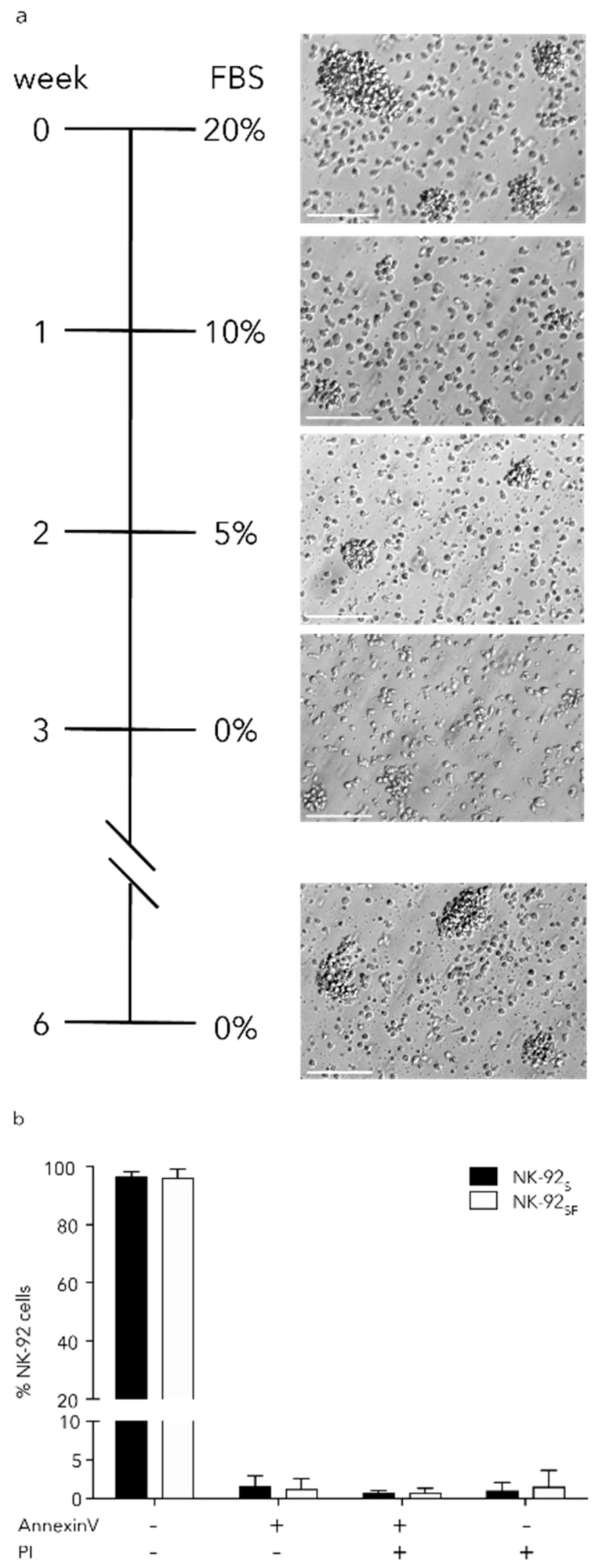
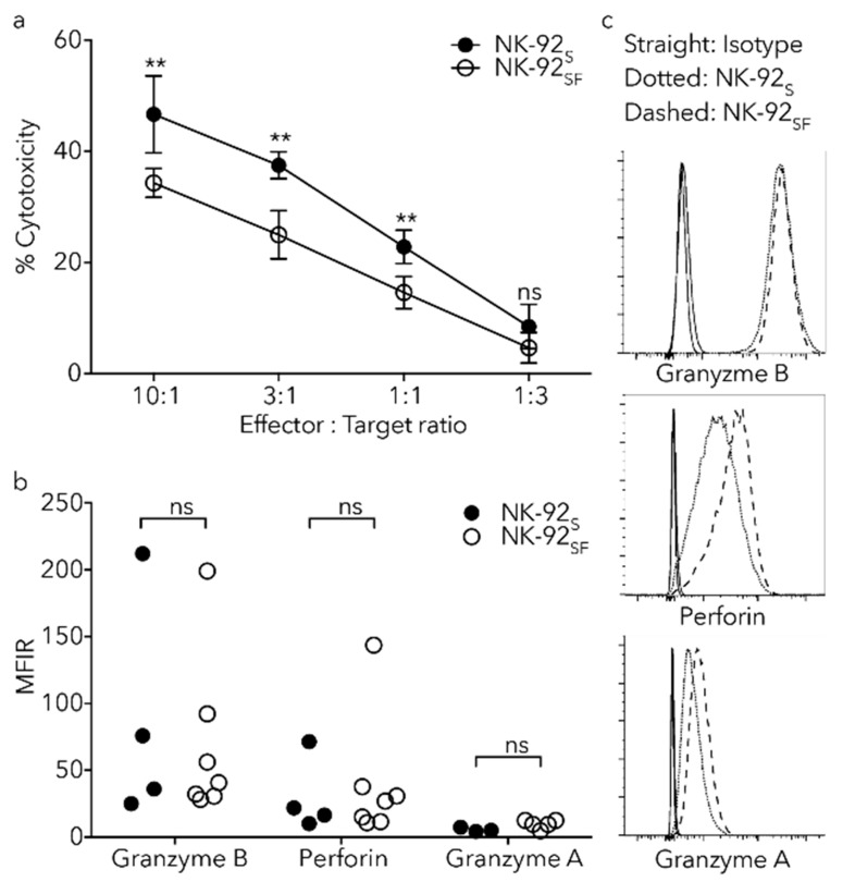
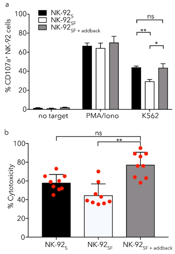
Natural killer (NK) cells stand out as promising candidates for cellular immunotherapy due to their capacity to kill malignant cells. However, the therapeutic use of NK cells is often dependent on cell expansion and activation with considerable amounts of serum and exogenous cytokines. We aimed to develop an expansion protocol for NK-92 cells in an effort to generate a cost-efficient, xeno-free, clinical grade manufactured master cell line for therapeutic applications. By making functional assays with NK-92 cells cultured under serum-free conditions (NK-92SF) and comparing to serum-supplemented NK-92 cells (NK-92S) we did not observe significant alterations in the viability, proliferation, receptor expression levels, or in perforin and granzyme levels. Interestingly, even though NK-92SF cells displayed decreased degranulation and cytotoxicity against tumor cells in vitro, the degranulation capacity was recovered after overnight incubation with 20% serum in the medium. Moreover, lentiviral vector-based genetic modification efficiency of NK-92SF cells was comparable with NK-92S cells. The application of similar strategies can be useful in reducing the costs of manufacturing cells for clinical use and can help us understand and implement strategies towards chemically defined expansion and genetic modification protocols.

Keywords: NK cell; NK-92; immunotherapy; serum-free.


Conflict of interest statement

The authors declare no conflict of interest.

Figures

Figure 1
Figure 1
Serum-free culture of NK-92 cells does not affect colony formation and viability. (a) Schematic overview of the serum starvation process over four weeks following a three-week recovery phase. Microscope images of NK-92 cells during the starvation process. Scale bar equals 100 μm. (b) Staining with Annexin V and propidium iodide (PI) of CD56+ NK-92 cells, after the 3-week recovery period, identified live cells (Annexin V/PI), pre- (Annexin V+/PI) and pro-apoptotic (Annexin V+/PI+) cells as well as necrotic cells (Annexin V/PI+). Each bar represents the mean (+SD) of eight independent assays performed in duplicates.
Figure 2
Figure 2
Proliferation of serum-free NK-92 cell culture is slightly reduced. (a) Staining of NK-92S/SF cells for KI-67 as a measurement of proliferation. Each bar represents the mean (+SD) of eight independent assays performed in duplicates. (b) Cell division between NK-92S and NK-92SF cells was monitored for three days. Analyzed by comparing intracellular carboxyfluorescein succinimidyl ester (CFSE) dilution to day 0. Each data point represents the mean (±SD) of two independent assays performed in triplicates. (c) Representative CFSE histograms for four consecutive days. (d) Cell division of NK-92 cells was monitored for 18 days after the recovery phase and fold expansion was calculated compared to day 0. The graph shows one representative experiment where each data point represents the mean (±SD) of triplicates. (e) Representative karyotyping picture from NK-92SF cells after the recovery phase. Arrowheads show previously published alterations of the chromosomes in NK-92 cells. Colors were inverted from the original image. (a): Welch’s t-test was used.
Figure 3
Figure 3
Receptor expression is not altered on NK-92SF cells. NK-92 cells were analyzed with a full panel of NK-cell markers. Only markers with detected expression, i.e., higher than unstained samples, are shown in the figure. Geometric mean fluorescent intensity (gMFI) of NK cell markers activation markers (a) or adhesion markers (c) on NK-92S and NK-92SF cells. (b) Representative histograms showing indicated receptor expression. Straight line shows isotype control, dotted line shows receptor expression on NK-92S, and dashed line shows receptor expression on NK-92SF. (d) Representative histograms for adhesion receptors on NK-92 cells. Straight line shows unstained control, dotted line shows receptor expression on NK-92S cells, and dashed line shows receptor expression on NK-92SF cells. Floating bars show mean and min/max value of three independent assays performed as a single or duplicate experiment.
Figure 4
Figure 4
Analysis of gene expression profiles in NK-92 cells exposed to different concentrations of serum. (a) Canonical pathway analysis for NK-92 cells exposed to 5% serum compared to cells grown without serum; (b) the heatmap shows statistically significant differentially regulated pathways created by comparison of differential gene expression contrasts for contrasts indicated below; (c) differential gene expression for pathways related to immune response. Missing values that did not reach statistical significance criteria are shown in black. (d) Ingenuity Pathway Analysis (IPA) upstream transcriptional regulator analysis for genes that are found to be differentially regulated in datasets.
Figure 5
Figure 5
Reduced degranulation rate and cytotoxicity against K562 in NK-92SF cells. (a) Chromium release assay, with K562 cells as target cells, show reduced cytotoxicity in NK-92SF cells at designated effector:target ratios. Each data point represents the mean (+SD) of two independent assays performed in triplicates. (b) Load of Perforin, Granzyme A, and Granzyme B in NK-92S and NK-92SF cells as shown by gMFI ratio to isotype do not alter between culture conditions. Each bar represents the mean (+SD) of four independent assays performed in single or duplicate experiments. (c) Representative histograms showing indicated marker expression. Straight line shows isotype control, dotted line shows receptor expression on NK-92S cells and dashed line shows receptor expression on NK-92SF cells. Statistical significance (** p < 0.01). (a): 2way ANOVA and (b): Wilcoxon test was used.
Figure 6
Figure 6
Addback of serum to NK-92SF cells recovers killing capacity. (a) NK-92SF show lower degranulation capacity against K562 target cells during a 5 h co-culture compared to NK-92S. NK-92SF cells were cultured in 20% serum-containing media for 16 h (NK-92SF+addback) and degranulation was tested against K562 target cell line. (b) Re-introduction of 20% serum for 16 h (NK-92SF+addback) increases killing capacity in a 4 h chromium release assay significantly. (c) NK-92SF cells are able to recover from freezing and thawing and show no altered viability 72 h post-thaw regardless of serum concentrations. Staining of CD56+ NK-92 cells with Annexin V and propidium iodide identified a proportion of live cells (Annexin V/PI), pre- (Annexin V+/PI) and pro-apoptotic (Annexin V+/PI+) cells, as well as dead cells (Annexin V/PI+). (d,e) NK-92 cells previously cultured with or without serum were kept in liquid nitrogen for long-term storage and then thawed and cultured for 16 h in complete medium (SCGM + 20% FBS). No significant difference in degranulation or cytotoxic capacity of NK-92SF compared to NK-92S cells could be observed. For (a,b) and (e) each graph represents the mean (+SEM) of three independent assays performed in triplicates. (c,d) each graph represents the mean (+SEM) of two independent assays performed in duplicates. Statistical significance (* p < 0.05; ** p < 0.01). (a,d): Welch’s t-test, (b): Kruskal–Wallis test, (e): Two-way ANOVA were used.
Figure 6
Figure 6
Addback of serum to NK-92SF cells recovers killing capacity. (a) NK-92SF show lower degranulation capacity against K562 target cells during a 5 h co-culture compared to NK-92S. NK-92SF cells were cultured in 20% serum-containing media for 16 h (NK-92SF+addback) and degranulation was tested against K562 target cell line. (b) Re-introduction of 20% serum for 16 h (NK-92SF+addback) increases killing capacity in a 4 h chromium release assay significantly. (c) NK-92SF cells are able to recover from freezing and thawing and show no altered viability 72 h post-thaw regardless of serum concentrations. Staining of CD56+ NK-92 cells with Annexin V and propidium iodide identified a proportion of live cells (Annexin V/PI), pre- (Annexin V+/PI) and pro-apoptotic (Annexin V+/PI+) cells, as well as dead cells (Annexin V/PI+). (d,e) NK-92 cells previously cultured with or without serum were kept in liquid nitrogen for long-term storage and then thawed and cultured for 16 h in complete medium (SCGM + 20% FBS). No significant difference in degranulation or cytotoxic capacity of NK-92SF compared to NK-92S cells could be observed. For (a,b) and (e) each graph represents the mean (+SEM) of three independent assays performed in triplicates. (c,d) each graph represents the mean (+SEM) of two independent assays performed in duplicates. Statistical significance (* p < 0.05; ** p < 0.01). (a,d): Welch’s t-test, (b): Kruskal–Wallis test, (e): Two-way ANOVA were used.
Figure 7
Figure 7
Serum-starvation of NK-92 cells does not change transduction efficiency with lentiviral vectors. CD56+GFP+ NK-92 cells three days after transduction with non-purified (a) or purified (b) LeGO-G2 virus in the absence or presence of BX-795. The graph represents the mean (+SEM) of two independent assays performed in duplicates. The LeGO-G2 virus is only introduced to express GFP, and the result was analyzed as GFP+ percentage three days post-transduction.

References

    1. Dahlberg C.I., Sarhan D., Chrobok M., Duru A.D., Alici E. Natural Killer Cell-Based Therapies Targeting Cancer: Possible Strategies to Gain and Sustain Anti-Tumor Activity. Front. Immunol. 2015;6:605. doi: 10.3389/fimmu.2015.00605. - DOI - PMC - PubMed
    1. Sutlu T., Alici E. Natural killer cell-based immunotherapy in cancer: Current insights and future prospects. J. Intern. Med. 2009;266:154–181. doi: 10.1111/j.1365-2796.2009.02121.x. - DOI - PubMed
    1. Cheng M., Ma J., Chen Y., Zhang J., Zhao W., Zhang J., Wei H., Ling B., Sun R., Tian Z. Establishment, characterization, and successful adaptive therapy against human tumors of NKG cell, a new human NK cell line. Cell Transplant. 2011;20:1731–1746. doi: 10.3727/096368911X580536. - DOI - PubMed
    1. Robertson M.J., Cochran K.J., Cameron C., Le J.M., Tantravahi R., Ritz J. Characterization of a cell line, NKL, derived from an aggressive human natural killer cell leukemia. Exp. Hematol. 1996;24:406–415. - PubMed
    1. Gong J.H., Maki G., Klingemann H.G. Characterization of a human cell line (NK-92) with phenotypical and functional characteristics of activated natural killer cells. Leukemia. 1994;8:652–658. - PubMed

LinkOut - more resources