

Where should Kirschner wires be placed when fixing patella fracture with modified tension-band wiring? A finite element analysis
- PMID: 30634995
- PMCID: PMC6329102
- DOI: 10.1186/s13018-019-1060-x
Where should Kirschner wires be placed when fixing patella fracture with modified tension-band wiring? A finite element analysis
Abstract
Background: The position of Kirschner wires (K-wires) has an influence on the outcome of modified tension-band wiring (MTBW) in fixing patella fractures. However, the instruction for K-wires positioning is not clear enough. This study tried to clarify the effect of K-wires positioning and provide evidence for a more definite instruction.
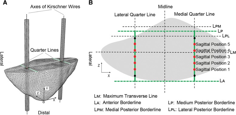
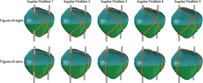
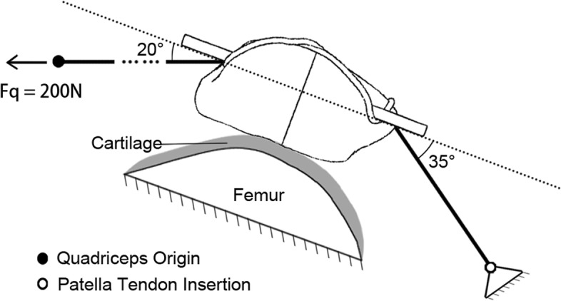
Methods: The sagittal position (SP) suitable for placing K-wires was evenly divided into SP 1-5 from anterior to posterior, and the finite element models of midpatella transverse fractures fixed by the figure-of-eight or figure-of-zero MTBW were built up at each SP. Separating displacement of the fracture, stress of the fracture, and stress of the internal fixations were measured at 45° knee flexion by using finite element analysis.
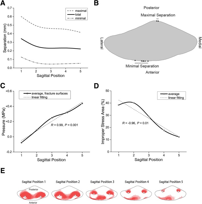
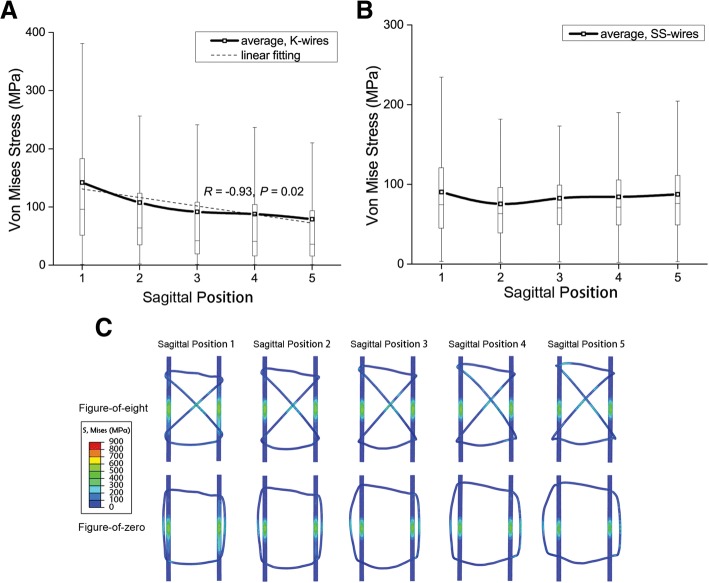
Results: The separating displacement of the fracture was smaller at SP 3-5 (23% smaller than SP 1-2). From SP 1 to 5, the compression of the fracture surfaces increased (R = 0.99, P = 0.001); the improper stress area of the fracture surfaces decreased (R = - 0.96, P = 0.01), and so was the stress of K-wires (R = - 0.93, P = 0.02). However, the stress of stainless steel wires showed a stable trend.
Conclusions: The SP of K-wires plays a role in the function of MTBW in the surgical management of transverse patella fracture. At 45° knee flexion, posteriorly placed (close to the articular surface) K-wires enable optimal stability and stress for the fracture, which provides basis for the positioning of K-wires in clinical practice.
Keywords: Biomechanics; Finite element analysis; Kirschner wire; Modified tension-band wiring; Patella fracture.
Conflict of interest statement
Ethics approval and consent to participate
CT image acquisition was approved by Ethics Committee of Shanghai Sixth People’s Hospital (Approval No. 2016-143) and a written consent was obtained from the participant.
Consent for publication
The participant enrolled into the study agreed the use of data for research.
Competing interests
The authors declare that they have no competing interests.
Publisher’s Note
Springer Nature remains neutral with regard to jurisdictional claims in published maps and institutional affiliations.
Figures
References
-
- Nummi J. Fracture of the patella. A clinical study of 707 patellar fractures. Ann Chir Gynaecol Fenn Suppl. 1971;179:1–85. - PubMed
-
- Bucholz RW, Heckman JD, Court-Brown CM. Fractures of the patella and injuries to the extensor mechanism. In: Harris RM, editor. Rockwood & Green’s fractures in adults. 6. Philadelphia: Lippincott Williams & Wilkins; 2006. pp. 1969–1997.
-
- Rüedi TP, Buckley RE, Moran CG. AO principles of fracture management. Stuttgart: Thieme; 2007.
MeSH terms
Grants and funding
LinkOut - more resources
Full Text Sources
Medical
