Review
. 2019:103:33-70.
doi: 10.1016/bs.aivir.2018.09.003. Epub 2018 Nov 28.

Phage Lysis: Multiple Genes for Multiple Barriers

Affiliations
Review

Phage Lysis: Multiple Genes for Multiple Barriers

Jesse Cahill et al. Adv Virus Res. 2019.

Abstract

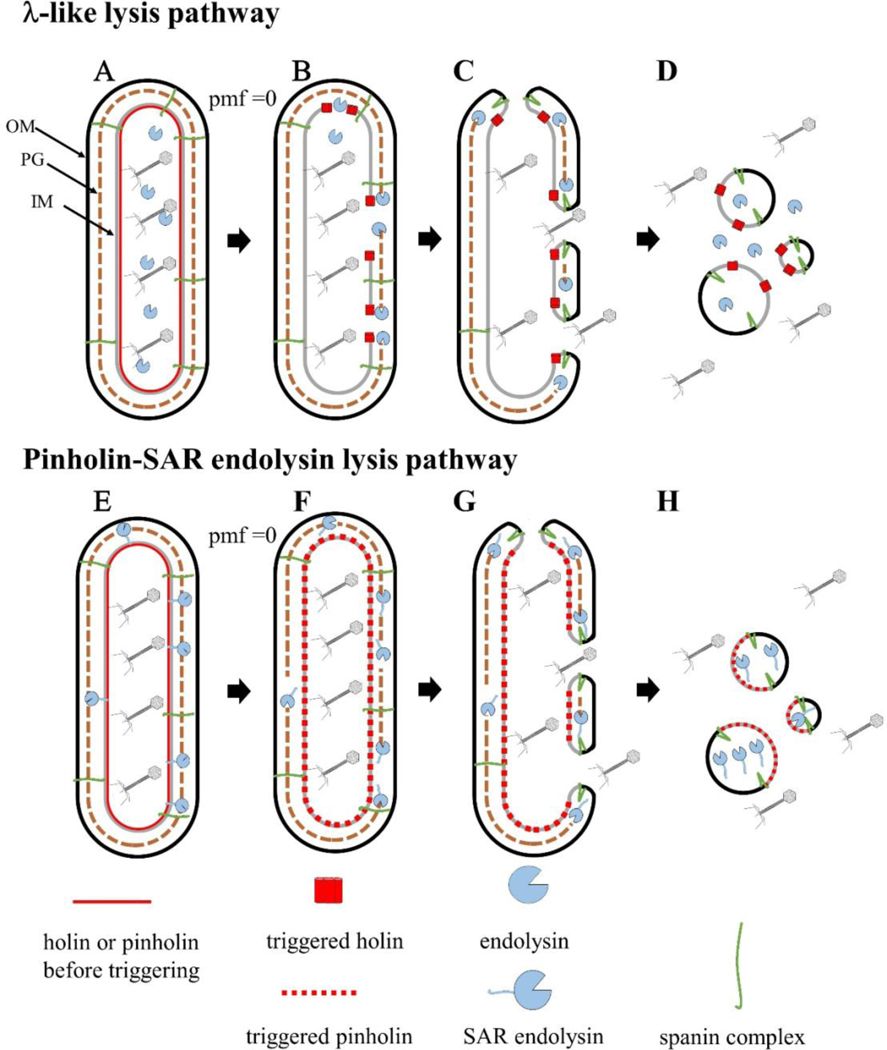
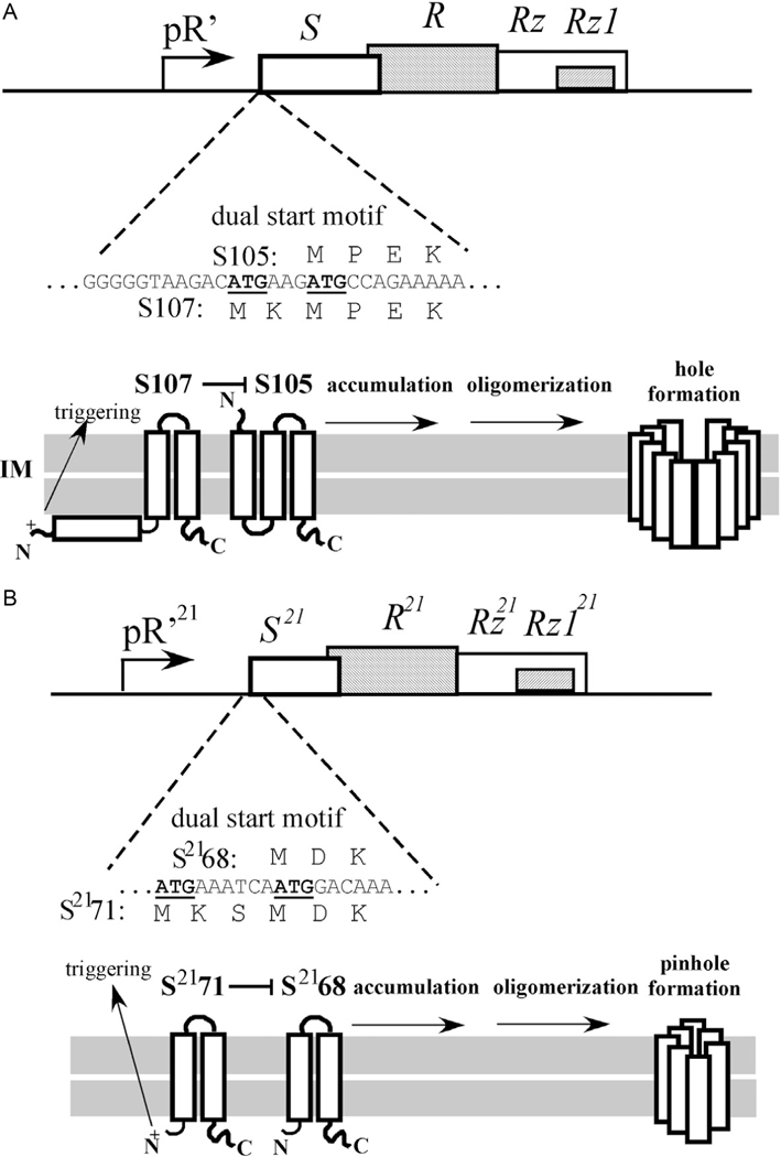
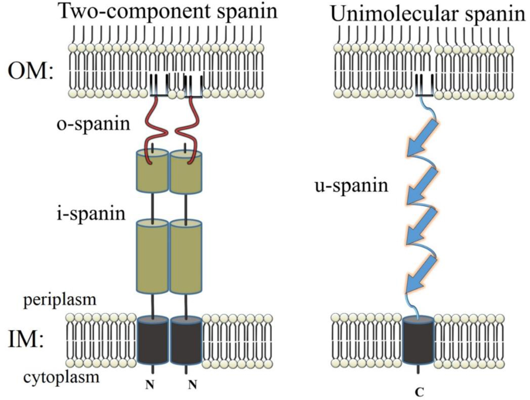
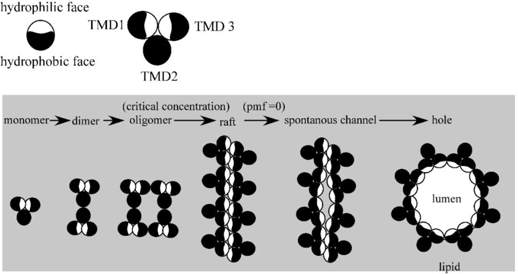
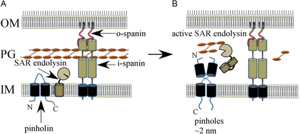
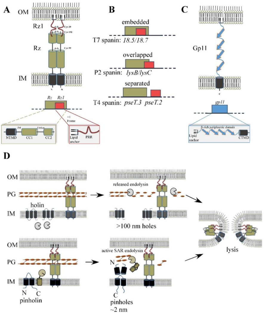
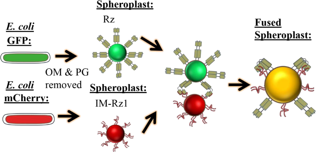
The first steps in phage lysis involve a temporally controlled permeabilization of the cytoplasmic membrane followed by enzymatic degradation of the peptidoglycan. For Caudovirales of Gram-negative hosts, there are two different systems: the holin-endolysin and pinholin-SAR endolysin pathways. In the former, lysis is initiated when the holin forms micron-scale holes in the inner membrane, releasing active endolysin into the periplasm to degrade the peptidoglycan. In the latter, lysis begins when the pinholin causes depolarization of the membrane, which activates the secreted SAR endolysin. Historically, the disruption of the first two barriers of the cell envelope was thought to be necessary and sufficient for lysis of Gram-negative hosts. However, recently a third functional class of lysis proteins, the spanins, has been shown to be required for outer membrane disruption. Spanins are so named because they form a protein bridge that connects both membranes. Most phages produce a two-component spanin complex, composed of an outer membrane lipoprotein (o-spanin) and an inner membrane protein (i-spanin) with a predominantly coiled-coil periplasmic domain. Some phages have a different type of spanin which spans the periplasm as a single molecule, by virtue of an N-terminal lipoprotein signal and a C-terminal transmembrane domain. Evidence is reviewed supporting a model in which the spanins function by fusing the inner membrane and outer membrane. Moreover, it is proposed that spanin function is inhibited by the meshwork of the peptidoglycan, thus coupling the spanin step to the first two steps mediated by the holin and endolysin.

Keywords: antiholin; endolysin; evolution; holin; lysis; multigene lysis; pinholin; single gene lysis; spanin.


Figures

Fig. 1
Fig. 1
Cartoon model of the two pathways of phage lysis of Gram-negative hosts. During the late morphogenesis period, the endolysin or SAR endolysin accumulates in the cytoplasm (A) or inner membrane (E), respectively. Upon reaching a critical concentration, holin triggering results in micron-scale holes (B) or small heptameric pinholes (F), which release the endolysin into the periplasm (B), or release the SAR end- olysin from the inner membrane into the periplasm;peptidoglycan degradation occurs at this step for both pathways. PG degradation results in spanin activation, which removes the topological barrier of the outer membrane by fusing both membranes (C and G). This results in the release of phage progeny and cytoplasmic content (D and H).
Fig. 2
Fig. 2
Comparison of two different holms with dual start motifs. (A) The lysis cassette of λ and the dual start motif of S (above). Below: The topology of S107 and S105. In this configuration, S107 negatively regulates S105. At the time of triggering the S107 TMD1 flips its N-terminus into the periplasm (see arrow). (B) The lysis cassette of phage 21 and the dual start motif of S21’ (above). Below: The topology of S2171 and S2168. In this configuration, S2171 negatively regulates S2168. At the time of triggering the S2171 TMD1 exits the bilayer and enters the periplasm (arrow).
Fig. 3
Fig. 3
Cartoon comparing topology of two-component spanins to u-spanins, the inner membrane (IM) and outer membrane (OM). The N-terminus of the i-spanin and the C-terminus of the o-spanin are labeled. The peptidoglycan is not depicted.
Fig. 4
Fig. 4
Model for the lambda holin function. A top-down view, each circle representing a TMD of S105. In the “death raft” model, the holin accumulates as dimers in which the more hydrophilic faces of TMD1 and TMD are sequestered intramolecularly. At a critical concentration, holin rafts form; here for simplicity, they are depicted as linear arrays, but the key feature is that in the rafts, many or all of the hydrophilic faces are now sequestered intermolecularly. Rafts cause local depolarization of the membrane, which in turn causes conformational changes in the holins leading to hole formation. The hydrophilic faces of TMD1 and TMD3 face the aqueous lumen of the hole (not drawn to scale).
Fig. 5
Fig. 5
Model for pinholin and SAR endolysin function. (A) The SAR endolysin accumulates in an inactive form, tethered to the IM. The TMD1 of the pinholin is in the IM. The N-terminal domain of the pinholin exits the bilayer gradually, which leads to the formation of dimers (B) that oligomerize (not shown) and trigger to form a functional pinholin. This causes the loss of the PMF, which results in the release of the SAR endolysin from the IM and its refolding to an enzymatically active form.
Fig. 6
Fig. 6
Spanin products, genes, and model for function. (A) Cartoon of λ spanin complex, comprised of Rz and Rz1, is shown within the cell envelope. The homotypic intermolecular disulfide linkages are denoted. Below: The Rz1gene is embedded within the +1 reading frame of Rz. Functional domains of the spanin complex that has been identified by genetic analysis are denoted in at relative positions respective to the predicted structural features. NTMD = N-terminal transmembrane domain. JM = juxtamembrane region, CC1 = alpha helix with predicted coiled-coil domains, CC2 = distal coiled-coil domain, PRR = proline-rich region. (B) Examples of different spanin genetic architectures, classified on the degree of overlap of the o-spanin gene (red) within the i-spanin (green). (C) The u-spanin gene product, exemplified by gp11 of T1. Below: Predicted domains of the gp11 product. (D) Model for spanin function following the canonical holin-endolysin (top) or pinholin-SAR endolysin pathway (bottom). For both pathways, spanins activate after PG degradation and undergo a conformational change that causes fusion of the IM and OM. This removes the topological barrier of the OM at the last step of lysis, which results in the release of phage progeny.
Fig. 7
Fig. 7
Spheroplast fusion assay. E. coli cells are induced for coexpression of GFP and Rz (green cell) or mCherry and Rz1 (red cell). Spheroplasts are formed by treating the cells with EDTA and lysozyme. IM-Rz1 is a mutant with an IM retention signal.
Fig. 8
Fig. 8
Model showing that phages with a shorter morphogenesis period have an advantage in a host-rich environment. Phages with late or early programmed lysis times are shown in black or red, respectively. The programmed lysis time for the red phage is time = 1 and time = 3 for the first and second generation, and the black phage lyses at time = 2. The late temporal program (A) has the advantage of releasing more phages in a single burst in the host-poor environment, compared to the shorter program (B). Below the dashed line: A host-rich environment will confer advantage to the early temporal program (C), since the cycles of exponential growth are shortened. Advantage is judged as the total free phage at time = 3, indicated as theoretical titer. It should be noted that for simplicity, variables such as the rate of adsorption or injection are not considered in this model.

References

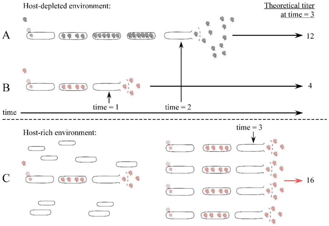
    1. Abedon ST, 1989. Selection for bacteriophage latent period length by bacterial density: a theoretical examination. Microb. Ecol. 18, 79. - PubMed
    1. Abedon ST, Herschler TD, Stopar D, 2001. Bacteriophage latent-period evolution as a response to resource availability. Appl. Environ. Microbiol. 67, 4233. - PMC - PubMed
    1. Barenboim M, Chang CY, dib Hajj F, Young R, 1999. Characterization of the dual start motif of a class II holin gene. Mol. Microbiol. 32, 715. - PubMed
    1. Bass RB, Strop P, Barclay M, Rees DC, 2002. Crystal structure of Escherichia coli MscS, a voltage-modulated and mechanosensitive channel. Science 298, 1582. - PubMed
    1. Beremand MN, Blumenthal T, 1979. Overlapping genes in RNA phage: a new protein implicated in lysis. Cell 18, 257. - PubMed

MeSH terms

Substances

LinkOut - more resources