Review
. 2019:342:27-71.
doi: 10.1016/bs.ircmb.2018.07.004. Epub 2018 Aug 29.

T Cells and Regulated Cell Death: Kill or Be Killed

Affiliations
Review

T Cells and Regulated Cell Death: Kill or Be Killed

Johan Spetz et al. Int Rev Cell Mol Biol. 2019.

Abstract

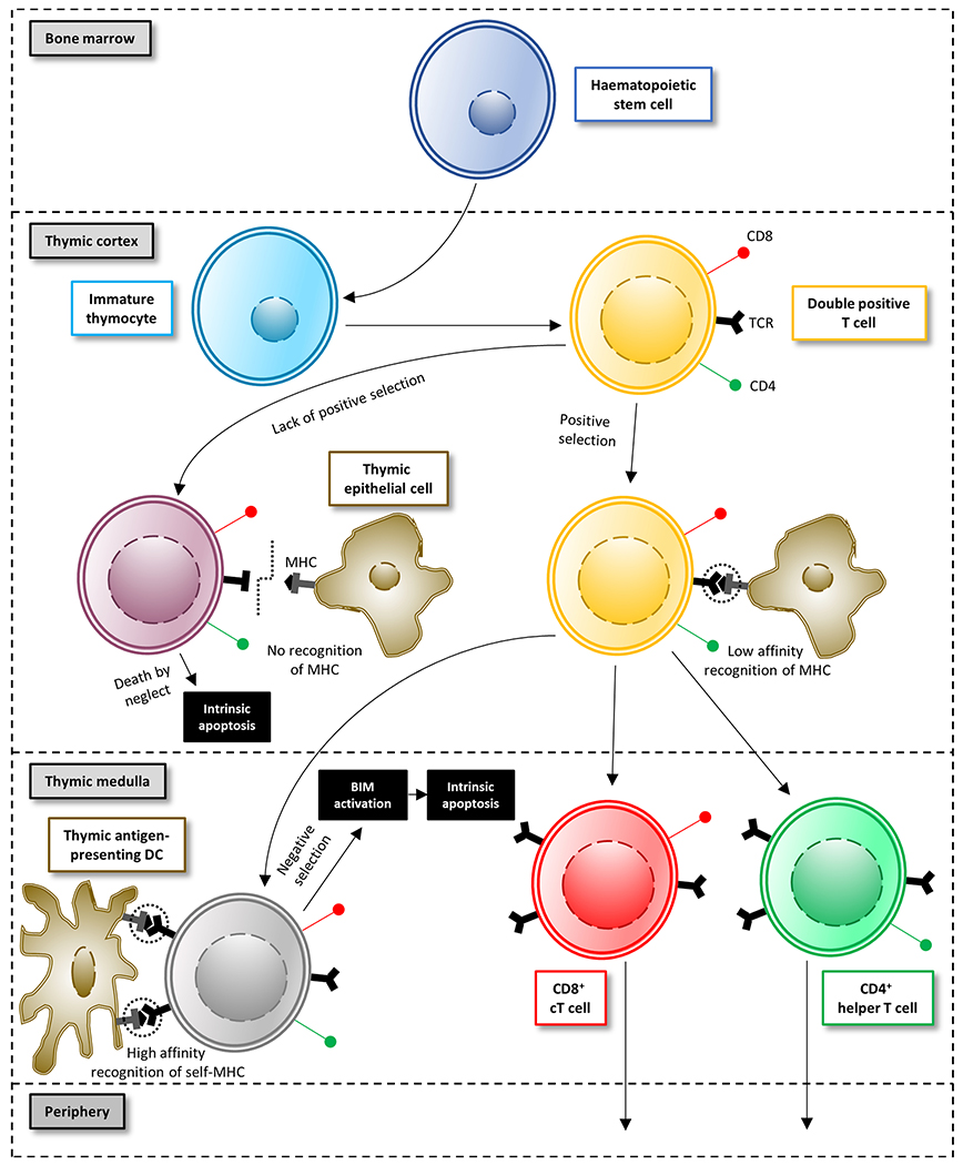
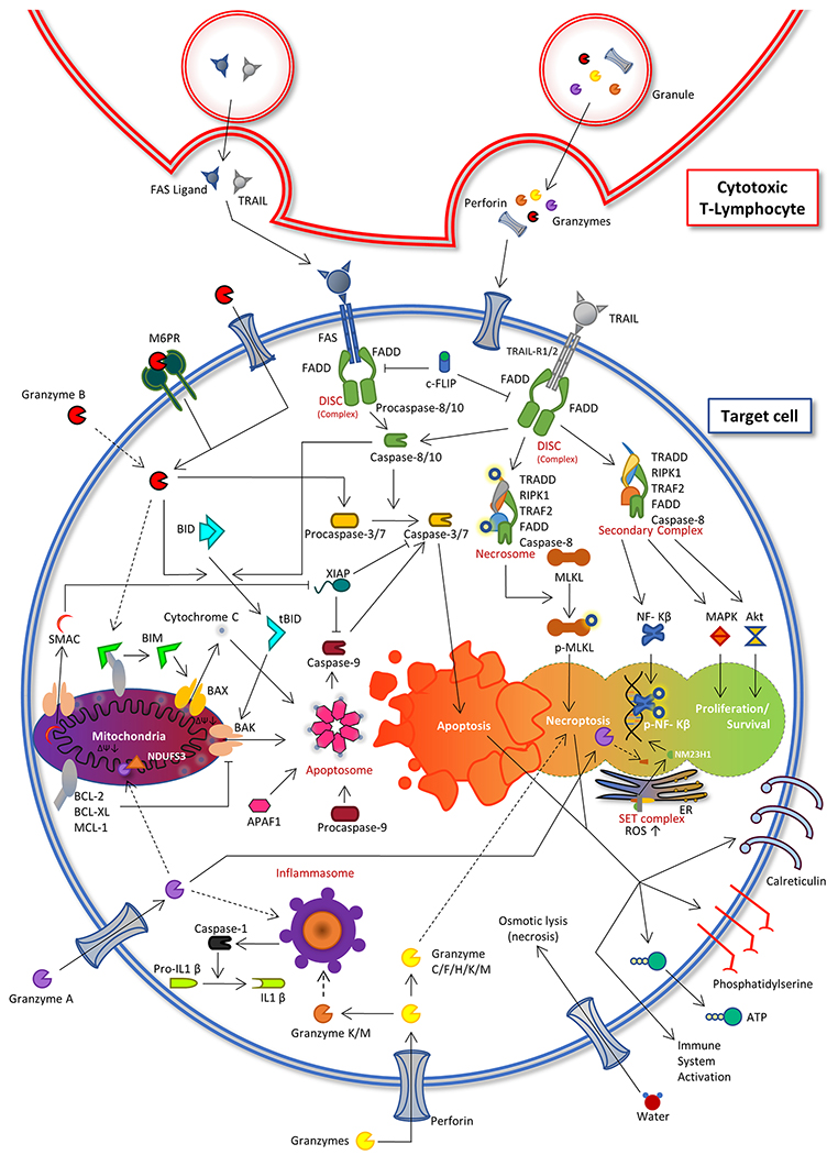
Cell death plays two major complementary roles in T cell biology: mediating the removal of cells that are targeted by T cells and the removal of T cells themselves. T cells serve as major actors in the adaptive immune response and function by selectively killing cells which are infected or dysfunctional. This feature is highly involved during homeostatic maintenance, and is relied upon and modulated in the context of cancer immunotherapy. The vital recognition and elimination of both autoreactive T cells and cells which are unable to recognize threats is a highly selective and regulated process. Moreover, detection of potential threats will result in the activation and expansion of T cells, which on resolution of the immune response will need to be eliminated. The culling of these T cells can be executed via a multitude of cell death pathways which are used in context-specific manners. Failure of these processes may result in an accumulation of misdirected or dysfunctional T cells, leading to complications such as autoimmunity or cancer. This review will focus on the role of cell death regulation in the maintenance of T cell homeostasis, as well as T cell-mediated elimination of infected or dysfunctional cells, and will summarize and discuss the current knowledge of the cellular mechanisms which are implicated in these processes.

Keywords: Apoptosis; Cancer immunotherapy; Cell death; Ferroptosis; Granzymes; Immunosurveillance; Necroptosis; Pyroptosis; T cell.


Figures

Figure 1:
Figure 1:. Maturation and selection of T cells.
T cells originate from haematopoietic stem cells, formed in the bone marrow. Haematopoietic progenitor cells are expanded into immature thymocytes (lacking CD4, CD8 and TCR) in the cortex of the thymus. These are developed into double-positive (CD4+, CD8+) thymocytes which express TCR with different affinity for different MHC. Double-positive thymocytes go through positive selection by exposure to self MHC–endogenous peptide complexes (expressed mainly on thymic epithelial cells). Thymocytes which recognise and interact with MHC receive survival signals from the thymic epithelial cells, while thymocytes with no recognition of MHC fail to receive the survival signals and are eliminated via apoptosis (“death by neglect”). The positively selected T cells migrate through the thymic medulla and undergo negative selection, where self-reactive T cells are culled. T cells (with CD4 or CD8 expression) which bind with high affinity to self-MHC complexes on thymic antigen-presenting cells are eliminated, while remaining CD4+ and CD8+ T cells are released into the periphery.
Figure 2:
Figure 2:. Mechanisms of T cell-mediated cell death.
cT cells can eliminate other cells via granule exocytosis and/or the expression and release of death ligands. Granule exocytosis entails release of e.g. granzymes and perforin (PRF1) toward the contact site of the target infected/dysfunctional cell. The granzymes then enter the cell and are capable of inducing cell death via multiple different pathways, with granzyme B-induced apoptosis being the most commonly observed. Death ligand secretion (of e.g. FAS ligand or TRAIL) is also a possible mode of elimination. In this case, cell death is most often induced via the extrinsic apoptotic pathway, although it may in some contexts lead to necroptosis.

Similar articles

Cited by

References

    1. Aachoui Y, Leaf IA, Hagar JA, Fontana MF, Campos CG, Zak DE, Tan MH, Cotter PA, Vance RE, Aderem A, Miao EA, 2013. Caspase-11 protects against bacteria that escape the vacuole. Science (80-. ). 339, 975–978. 10.1126/science.1230751 - DOI - PMC - PubMed
    1. Abdool K, Cretney E, Brooks AD, Kelly JM, Swann J, Shanker A, Bere EW Jr., Yokoyama WM, Ortaldo JR, Smyth MJ, Sayers TJ, 2006. NK cells use NKG2D to recognize a mouse renal cancer (Renca), yet require intercellular adhesion molecule-1 expression on the tumor cells for optimal perforin-dependent effector function. J Immunol 177, 2575–2583. https://doi.org/177/4/2575 [pii] - PubMed
    1. Afshar-Sterle S, Zotos D, Bernard NJ, Scherger AK, Rödling L, Alsop AE, Walker J, Masson F, Belz GT, Corcoran LM, O’reilly LA, Strasser A, Smyth MJ, Johnstone R, Tarlinton DM, Nutt SL, Kallies A, 2014. Fas ligand-mediated immune surveillance by T cells is essential for the control of spontaneous B cell lymphomas. Nat. Med 20, 283–290. 10.1038/nm.3442 - DOI - PubMed
    1. Aglietti RA, Estevez A, Gupta A, Ramirez MG, Liu PS, Kayagaki N, Ciferri C, Dixit VM, Dueber EC, 2016. GsdmD p30 elicited by caspase-11 during pyroptosis forms pores in membranes. Proc. Natl. Acad. Sci 113, 7858–7863. 10.1073/pnas.1607769113 - DOI - PMC - PubMed
    1. Allard B, Pommey S, Smyth MJ, Stagg J, 2013. Targeting CD73 enhances the antitumor activity of anti-PD-1 and anti-CTLA-4 mAbs. Clin. Cancer Res. 19, 5626–5635. 10.1158/1078-0432.CCR-13-0545 - DOI - PubMed

Publication types

LinkOut - more resources