Comparative Study
. 2019 Jan 15;50(1):51-63.e5.
doi: 10.1016/j.immuni.2018.12.013. Epub 2019 Jan 8.

Oligoadenylate-Synthetase-Family Protein OASL Inhibits Activity of the DNA Sensor cGAS during DNA Virus Infection to Limit Interferon Production

Affiliations
Comparative Study

Oligoadenylate-Synthetase-Family Protein OASL Inhibits Activity of the DNA Sensor cGAS during DNA Virus Infection to Limit Interferon Production

Arundhati Ghosh et al. Immunity. .

Abstract

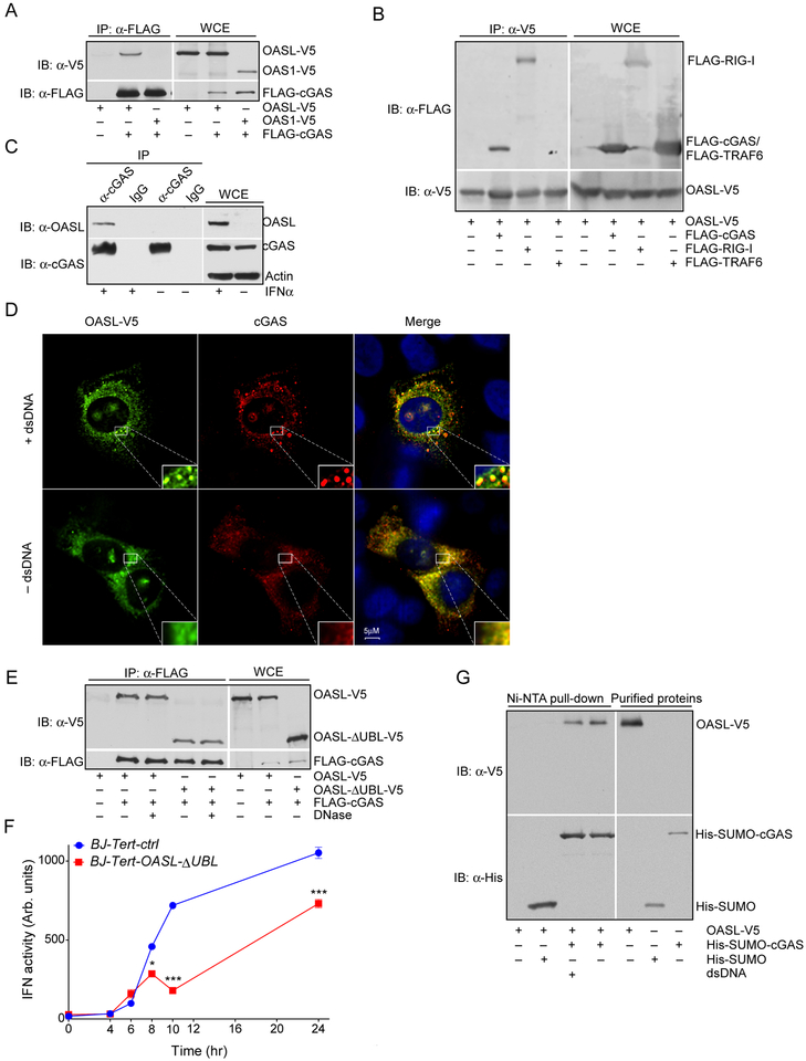
Interferon-inducible human oligoadenylate synthetase-like (OASL) and its mouse ortholog, Oasl2, enhance RNA-sensor RIG-I-mediated type I interferon (IFN) induction and inhibit RNA virus replication. Here, we show that OASL and Oasl2 have the opposite effect in the context of DNA virus infection. In Oasl2-/- mice and OASL-deficient human cells, DNA viruses such as vaccinia, herpes simplex, and adenovirus induced increased IFN production, which resulted in reduced virus replication and pathology. Correspondingly, ectopic expression of OASL in human cells inhibited IFN induction through the cGAS-STING DNA-sensing pathway. cGAS was necessary for the reduced DNA virus replication observed in OASL-deficient cells. OASL directly and specifically bound to cGAS independently of double-stranded DNA, resulting in a non-competitive inhibition of the second messenger cyclic GMP-AMP production. Our findings define distinct mechanisms by which OASL differentially regulates host IFN responses during RNA and DNA virus infection and identify OASL as a negative-feedback regulator of cGAS.

Keywords: DNA virus; IFN; OASL; cGAMP; cGAS; transcriptional signalling.


Conflict of interest statement

DECLARATION OF INTEREST

PS and SHT are full time employees and may have ownership interests at Western Oncolytics at present. However, the work presented here were carried out while they were at the University of Pittsburgh and no part of this work was carried out at the company.

Figures

Fig. 1:
Fig. 1:. RNA and DNA virus growth and pathogenesis in Oasl2−/− mice.
(A) Four to six weeks old WT and Oasl2−/− mice (N=9 each) were infected intranasally with VSV-GFP (2 × 10⁵ pfu/mice). Representative immunohistochemistry of 4 days post-infection (dpi) mouse brain sections stained with GFP antibody are shown.. (B) Olfactory bulbs were harvested from WT and Oasl2−/− mice at indicated days. VSV transcripts were quantified from the tissue homogenate by RT-qPCR. (C),(D) and (E) Four to six weeks old WT and Oasl2−/− mice (N=8 each) were infected intranasally with VV-Luc (2 × 10⁴ pfu/mice). Virus replication and spread were quantified on indicated days by whole animal bioluminescence imaging. (F) Losses of body weight of the mice infected as above were measured at indicated days postinfection. Results shown are pooled samples from twice repeated in vivo infection studies. For each data point mean and SEM were plotted, statistical significances were calculated by two-way ANOVA with Sidak’s multiple comparison test
Fig. 2:
Fig. 2:. Replication of multiple DNA viruses in OASL or Oasl2 deficient cells.
(A) and (B) Tail fibroblasts from WT and Oasl2−/− mice (A), and human BJ-Tert fibroblasts (B) were infected with VV-Luc (IHD-J strain) at MOI of 1. Virus replication was measured in triplicates at indicated times post-infection (hpi: hrs post-infection) by luciferase activity assay using IVIS200. (C) WT and OASL-KO BJ-Tert cells were infected with VV as above. Infectious virus particles in the culture media were collected and quantified by plaque assay on BSC-1 cells. (D)Equal numbers of WT and OASL-KO BJ-Tert cells were infected with HSV-1 at MOI of 1, 0.5 and 0.1. Culture supernatant were collected at 48 h (MOI of 1 and 0.5) and 72 h (MOI of 0.1) post-infection and infectious particles were quantified by plaque assay on Vero cells. (E) and (F) Equal numbers of fibroblasts from WT and Oasl2−/− mice were infected with HSV at MOI of 0.1 for 72 h (E), and with MCMV at MOI of 0.1 for 48 h (F). Infectious particles in the culture supernatant were measured by plaque assay on Vero cells and NIH3T3 cells respectively. (G) WT and OASL-KO BJ-Tert cells were infected with Adeno-GFP at MOI as indicated. Following 36 h of infection virus replication were quantified by measuring percentage of GFP+ cells by flow cytometry. Results shown are pooled samples from three times repeated in vitro infection studies. For each data point mean and SEM were plotted. Statistical significance were calculated by two-way ANOVA with Sidak’s multiple comparison test or by two-tailed Student’s t-test (E and (F).
Fig. 3:
Fig. 3:. Effect of OASL and Oasl2 on IFN induction by RNA and DNA viruses.
(A) WT and Oasl2−/− mice were infected with VV (IHD-J strain) as before (Fig. 1C). Mice were sacrificed as indicated followed by the collection of total cells from the BAL fluid of each mice, and quantitation of Ifnα mRNA by RT-qPCR. (B) Equal numbers of WT and OASL-KO BJ-Tert cells were infected with VV at a MOI of 1. Total RNA was collected at 12 and 24 hpi, followed by the quantitation of IFNβ, IFNα and IFIT1 mRNA by RT-qPCR. (C) Indicated cells were infected with HSV at MOI of 1. IFNβ mRNA induction was measured at different time points. (D) WT and OASL-KO BJ-Tert cells were infected with HSV-d109 for 24 h with indicated doses followed by the analysis of IFNβ mRNA induction. (E) Indicated cells were infected with HSV-d109 at MOI of 1 followed by the analysis of IFN03B2 and OASL mRNA induction by RT-qPCR. (F) WT and OASL-KO BJ-Tert cells were infected with HSV-d109 at 2 MOI for the indicated times followed by the RNAseq analysis of the poly(A) containing transcriptome. Heatmap of a subset of genes that are significantly upregulated in OASL-KO cells is shown. (G) BJ-Tert cells stably transduced with lentivirus carrying OASL or control vector were infected with HSV-d109 at MOI of 10 and 20 for 24 h. Culture supernatants were analyzed for IFNβ protein production after 24 h post-infection by ELISA. Results shown are representative of at least three times repeated in vitro experiments (B) to (E) and (G). In panel (A), pooled samples from twice repeated in vivo infection studies were plotted, while the average values of twice repeated RNAseq experiments were used for the heatmap in (F).
Fig. 4:
Fig. 4:. Characterization of OASL effect on DNA-sensor signaling pathway.
(A) THP1 cells transduced similarly as before with OASL-expressing lentivirus were stimulated with dsDNA by transfection with 100 bp synthetic DNA (1 μg/ml) for the indicated times. Culture supernatants were analyzed for IFNβ protein production by ELISA. (B) BJ-Tert OASL-KO cells were transduced with lentivirus carrying OASL followed by dsDNA transfection as indicated. IFNβ induction was measured by RT-qPCR. (C) Indicated cells were treated overnight with 1000 U of IFNα to induce OASL followed by dsDNA transfection (100 bp synthetic DNA, 1 μg/ml) for the indicated times. Whole cell lysates were analyzed for IRF3 phosphorylation by immunoblotting with Phospho-IRF-3 (Ser396) antibody along with total IRF3 and Actin antibodies. (D) WT and OASL-KO THP1 cells were stimulated either with dsDNA (100 bp synthetic DNA, 1 μg/ml) or with cGAMP (5 μg/ml) by transfection. IFNβ mRNA induction was analyzed after 6 h post-transfection by RT-qPCR. (E) WT and OASL-KO BJ-Tert cells were permeabilized with Digitonin and treated with cGAMP (5 μg/ml) for 4 and 6 hrs as indicated. Cell lysates were immunoblotted with ISG60 (IFIT3), OASL and actin antibody. (F) WT and cGAS-deficient (cGAS-KO) BJ-Tert cells were transduced with lentivirus carrying either OASL or the control vector and validated for protein expression by immunoblotting (inset). Cells were then infected with VV-Luc at MOI of 1 and the virus replication measured at indicated times by luciferase assays. Results shown are representative of at least three times repeated in vitro experiments.
Fig. 5:
Fig. 5:. Interaction of OASL and cGAS.
(A) C-terminal V5-tagged OASL (OASL-V5) or OAS1 (OAS1-V5) were cotransfected with Nterminal FLAG-tagged cGAS (FLAG-cGAS) in HEK293 cells for 24 h as indicated. Equal amounts of cell extracts (750 μg total protein) were immuneprecipitated (IP) with FLAG antibody (mouse monoclonal) followed by immunoblotting (IB) with either V5 (rabbit monoclonal) or FLAG (rabbit polyclonal) antibody. Whole cell lysates (WCE) were also immunoblotted in parallel to detect recombinant protein expression. (B) Similarly, OASL-V5 was cotransfected either with FLAG-cGAS or FLAG-TRAF6 followed by IP with V5 (mouse monoclonal) antibody and IB with FLAG or V5 antibody. (C) Control or IFNα treated HeLa cell extracts were immunoprecipitated either with cGAS antibody or control IgG followed by the detection of OASL with OASL antibody. Concomitant immunoblotting of the whole cell lysate shows the respective protein expression. (D) Immunofluorescence analysis of the OASL-V5 expressing HeLa cells with or without dsDNA transfection. HeLa cells stably transduced with OASL-V5 expressing lentivirus were stimulated with dsDNA by transfection for 4 h. Fixed cells were immunostained with cGAS and V5 antibodies followed by confocal microscopy. The insets show enlarged sections highlighting cGAS puncta formation after dsDNA stimulation. Scale bar = 5 μm. (E) OASL-V5 or OASL-ΔUBL-V5 constructs were cotransfected with FLAG-cGAS as indicated in HEK293 cells followed by IP as above. Immunoprecipitated samples were resuspended in DNase reaction buffer with or without DNase I as indicated, incubated at room temperature for 15 minutes followed by washing and analysis by IB. (F) BJ-Tert cells transduced with either control vector or OASL-ΔUBL-V5 carrying lentivirus were stimulated with dsDNA (100 bp synthetic DNA, 1 μg/ml) for the indicated times. IFN induction in the culture supernatant were assayed by IFN bioassay. (G) Purified OASL-V5 were mixed and incubated with either control His-SUMO or His-SUMOcGAS proteins followed by Ni-NTA pull-down in presence or absence of (100 bp synthetic DNA, 1 μg/ml). Pulled-down proteins were detected by IB as indicated. Representative results are shown from at least three times repeated experiments.
Fig. 6:
Fig. 6:. Influence of OASL on cGAMP production.
(A) Indicated cells were stimulated with dsDNA (100 bp synthetic DNA, 1 μg/ml) for 16 h followed by cGAMP quantitation in cell lysates by mass spectroscopy. (B) Purified OASL or buffer were mixed with purified cGAS proteins at indicated molar ratios and incubated on ice for 15 min followed by cGAS activity measurements by mass spectroscopy. (C) and (D) Non-competitive inhibition of cGAS activity by OASL. Purified cGAS and OASL proteins were mixed at 1:1 molar ratio followed by cGAS activity assays either in presence of constant GTP (C) or ATP (D). For each experiment cumulative results from three biological repeats are shown.

Similar articles

Cited by

References

    1. Ablasser A, Schmid-Burgk J, Hemmerling I, Horvath G, Schmidt T, Latz E, and Hornung V (2013). Cell intrinsic immunity spreads to bystander cells via the intercellular transfer of cGAMP. Nature 503, 530–534. - PMC - PubMed
    1. Aguirre S, and Fernandez-Sesma A (2017). Collateral Damage during Dengue Virus Infection: Making Sense of DNA by cGAS. J. Virol 91, e01081–16. - PMC - PubMed
    1. Andreeva L, Hiller B, Kostrewa D, Lässig C, de Oliveira Mann CC, Jan Drexler D, Maiser A, Gaidt M, Leonhardt H, Hornung V, et al. (2017). cGAS senses long and HMGB/TFAM-bound U-turn DNA by forming protein–DNA ladders. Nature 549, 394–398. - PubMed
    1. Auerbuch V, Brockstedt DG, Meyer-Morse N, O’Riordan M, and Portnoy DA (2004). Mice lacking the type I interferon receptor are resistant to Listeria monocytogenes. J. Exp. Med 200, 527–533. - PMC - PubMed
    1. Banerjee I, Behl B, Mendonca M, Shrivastava G, Russo AJ, Menoret A, Ghosh A, Vella AT, Vanaja SK, Sarkar SN, et al. (2018). Gasdermin D Restrains Type I Interferon Response to Cytosolic DNA by Disrupting Ionic Homeostasis. Immunity 49, 413–426.e5. - PMC - PubMed

Publication types

MeSH terms