. 2019 Jan 18;146(2):dev168963.
doi: 10.1242/dev.168963.

IMP regulates Kuzbanian to control the timing of Notch signalling in Drosophila follicle cells

Affiliations

IMP regulates Kuzbanian to control the timing of Notch signalling in Drosophila follicle cells

Weronika Fic et al. Development. .

Abstract

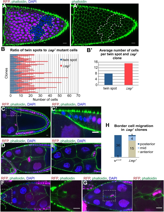
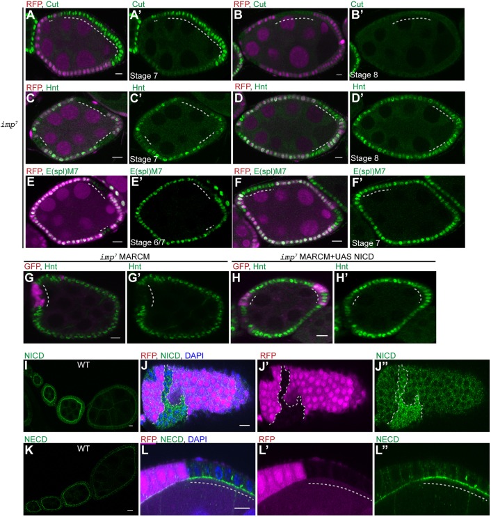
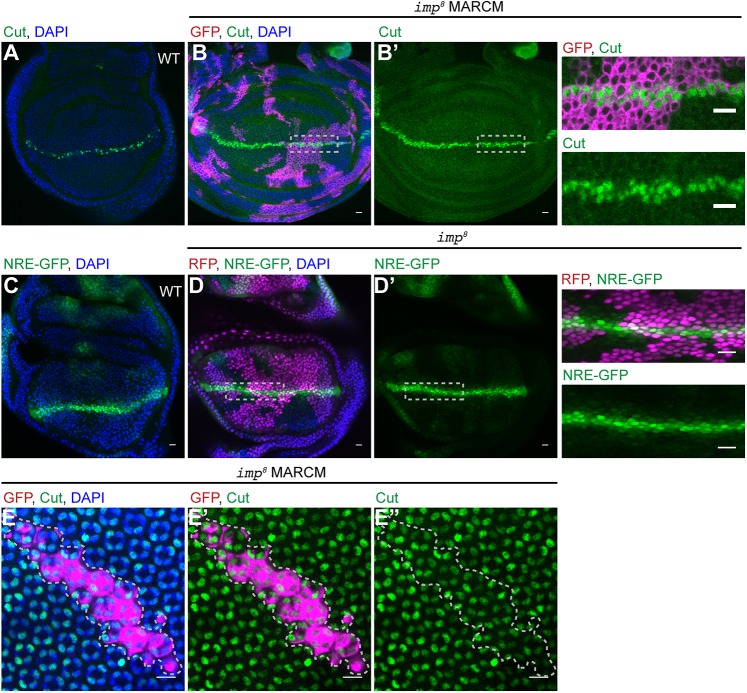
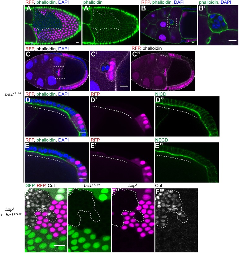
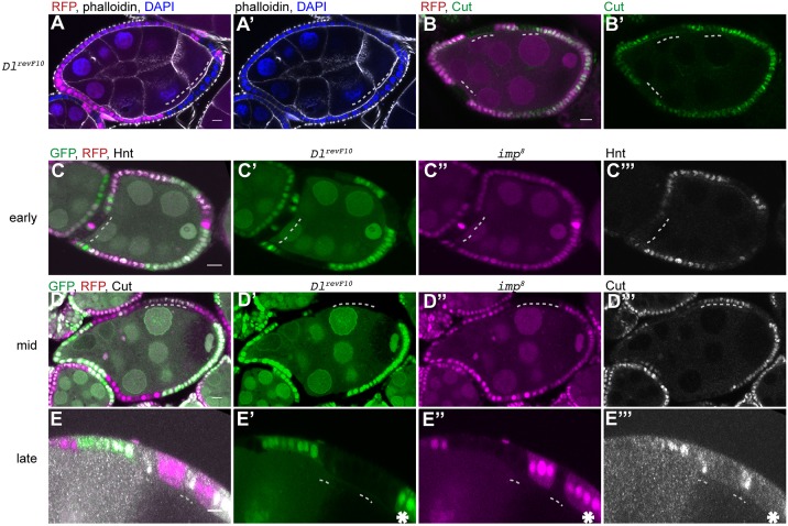
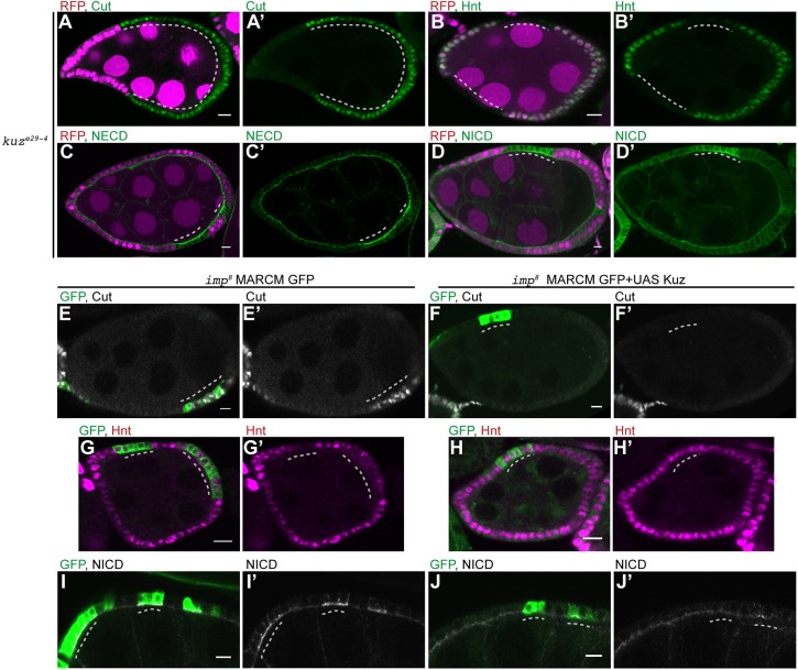
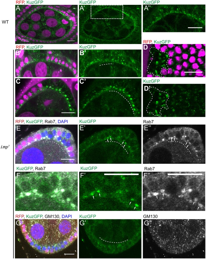
The timing of Drosophila egg chamber development is controlled by a germline Delta signal that activates Notch in the follicle cells to induce them to cease proliferation and differentiate. Here, we report that follicle cells lacking the RNA-binding protein IMP go through one extra division owing to a delay in the Delta-dependent S2 cleavage of Notch. The timing of Notch activation has previously been shown to be controlled by cis-inhibition by Delta in the follicle cells, which is relieved when the miRNA pathway represses Delta expression. imp mutants are epistatic to Delta mutants and give an additive phenotype with belle and Dicer-1 mutants, indicating that IMP functions independently of both cis-inhibition and the miRNA pathway. We find that the imp phenotype is rescued by overexpression of Kuzbanian, the metalloprotease that mediates the Notch S2 cleavage. Furthermore, Kuzbanian is not enriched at the apical membrane in imp mutants, accumulating instead in late endosomes. Thus, IMP regulates Notch signalling by controlling the localisation of Kuzbanian to the apical domain, where Notch cleavage occurs, revealing a novel regulatory step in the Notch pathway.

Keywords: ADAM10 protease; Delta; Drosophila oogenesis; RNA-binding protein.


Conflict of interest statement

Competing interestsThe authors declare no competing or financial interests.

Figures

Fig. 1.
Fig. 1.
imp mutant cells go through one extra division. (A) Surface view of a stage 10a egg chamber containing an imp7 mutant follicle cell clone (marked by the loss of RFP, magenta) stained with phalloidin (green) and DAPI (blue). The mutant cells are outlined in white. (B) Graph showing the number of wild-type and imp7 mutant cells in 56 independent twin spot clones. (B′) Histogram showing the average number of cells per wild-type clone and imp7 clone (n=56). (C,C′) Stage 10a chamber with an imp7 follicle cell clone that has not yet migrated posteriorly to envelop the oocyte (n=23). C′ shows magnification of the boxed area in C. Dashed line indicates mutant cells. (D,E) Wild-type stage 9 (n=12) (D) and stage 10b (n=21) (E) egg chambers showing the migration of the border cells between the nurse cells to reach the anterior of the oocyte at stage 10b. (F) Stage 9 egg chamber with an imp7 mutant clone that includes all of the border cells, which have failed to detach from the anterior (n=15). Image to the right is a magnification of the boxed area on the left. Dashed line indicates the border cell cluster. (G) Stage 9 egg chamber containing a mosaic of imp7 mutant and wild-type border cells. The mutant cells are found at the back of the cluster and trail behind the wild-type cells. The migration of these clusters is severely delayed and they often move only half way to the oocyte. Image to the right is a magnification of the boxed area on the left. (H) Quantification of the region to which wild-type border cell clusters and entirely mutant clusters have moved by stage 10b. Scale bars: 10 μm.
Fig. 2.
Fig. 2.
IMP is required for the first cleavage of Notch. (A,A′) Cut expression in a stage 7 egg chamber containing an imp7 follicle cell clone marked by the loss of RFP (magenta). Cut expression persists in the mutant cells (n=32). (B,B′) A stage 8 egg chamber containing an imp7 follicle cell clone stained for Cut (n=41). Cut is lost from the majority of mutant cells. (C,C′) Hnt expression in a stage 7 egg chamber containing several imp7 follicle cell clones marked by the loss of RFP. Hnt is expressed in the wild-type cells, but not in the mutant cells at stage 7 (n=28). (D,D′) A stage 8 egg chamber containing imp7 follicle cell clones stained for Hnt. The mutant cells have turned on Hnt at this stage (n=47). (E-F′) E(spl)m7lacZ expression in stage 6/7 and stage 7 egg chambers containing imp7 follicle cell clones marked by the loss of RFP. E(spl)m7lacZ is expressed at much lower levels in imp mutant cells than in wild-type cells at stage 6 (n=12), but at similar levels at stage 7 (n=21). (G,G′) A stage 7 egg chamber containing an imp7 MARCM clone marked by the expression of GFP (magenta) and stained for Hnt (green). The mutant cells do not express Hnt at stage 7 (n=27). (H,H′) A stage 7 egg chamber containing an imp7 MARCM clone expressing UAS-NICD stained for Hnt (green). Hnt expression is rescued by the expression of the NICD (n=21). (I-L″) Localisation of the NICD (I-J″) and the NECD (K-L″) in wild-type early-stage egg chambers and egg chambers containing imp mutant clones. Both NICD and NECD are enriched at the apical side of wild-type follicle cells until stage 6 when they are downregulated as a result of Delta signalling. NICD (J-J″; n=38) and NECD (L-L″; n=27) are not downregulated in imp7 mutant cells (marked by the loss of RFP). Dashed lines indicate imp mutant clones. Scale bars: 10 μm.
Fig. 3.
Fig. 3.
IMP is not a general component of the Notch signalling pathway. (A-B′) Third larval instar wing imaginal discs stained for Cut (green). B shows a disc containing imp8 MARCM clones marked by GFP expression (magenta). Cut is expressed normally in mutant cells along the dorsal-ventral compartment boundary (n=49). Images to the right are magnifications of the boxed areas in B and B′. (C) Third larval instar wing imaginal disc expressing the NRE-GFP reporter. (D,D′) A wing disc containing imp8 clones at the dorsal ventral boundary. NRE-GFP is expressed at similar levels in imp8 mutant and wild-type cells (n=16). Images to the right are magnifications of the boxed areas in D and D′. (E-E″) 50 h pupal eye disc containing imp8 MARCM clones marked by GFP expression (magenta). Clone area is outlined by dashed lines. Mutant cone cells are indistinguishable from wild type and express the cone cell marker Cut (green; n=19). Scale bars: 10 μm.
Fig. 4.
Fig. 4.
IMP does not act through the microRNA pathway. (A-C″) Stage 10a egg chambers containing belle47110 mutant clones marked by the loss of RFP. The mutant cells show a similar phenotype to that observed in imp mutants. They go through one extra round of division and are therefore smaller than the wild-type cells (A,A′; n=19). When all of the border cells are mutant for belle, there is a delay in border cell migration (B,B′; n=11). In mosaic border cell clusters, the mutant cells lag behind the wild-type border cells (C-C″). The migration of mutant follicle cells to envelop the oocyte is also delayed (white dashed line in C″; n=17). B′ and C′ are magnifications of the boxed areas in B and C, respectively. (D-E″) Stage 9 egg chambers containing belle47110 mutant clones marked by the loss of RFP stained for NICD (D-D″) and NECD (E-E″). The mutant cells retain high levels of NICD (D-D″; n=15) and NECD (E-E″; n=13) at their apical membranes. Dashed lines indicate mutant cells. (F-F‴) A stage 9 egg chamber containing both imp7 mutant clones marked by the loss of RFP (magenta) and belle47110 mutant clones marked by the loss of GFP (green), stained for Cut (white). Cut is still expressed in the double-mutant cells (marked by the dashed line), but not in the single-mutant cells (n=24). Scale bars: 10 μm.
Fig. 5.
Fig. 5.
imp is epistatic to Delta in follicle cells. (A-B′) Stage 6 egg chambers containing DlrevF10 mutant cells marked by the loss of RFP. The mutant cells are larger than wild type and have bigger nuclei, indicating that they have undergone the switch from mitosis to endoreplication prematurely (A,A′; n=18). The Dl mutant cells switch off Cut expression (green) before the wild-type cells (B,B′; n=17). Dashed lines indicate mutant cells. (C-E‴) Egg chambers containing both DlrevF10 clones marked by the loss of GFP (green) and imp8 mutant clones marked by the loss of RFP (magenta). (C-C‴) imp Dl double-mutant cells do not express Hnt (white) at stage 6/7, whereas Dl mutant cells do (n=34). (D-D‴) imp Dl double mutant cells still express Cut (white) at stage 6, unlike Dl mutant cells (n=56). (E-E‴) imp Dl double-mutant cells (marked by the dashed lines) still express Cut at stage 7, in contrast to wild-type cells (marked with an asterisk; n=23). Scale bars: 10 μm.
Fig. 6.
Fig. 6.
Kuzbanian is required for Notch activation in the follicle cells. (A-D′) Egg chambers containing kuze29-4 mutant cells marked by the loss of RFP. The kuz mutant cells continue to express Cut at stage 7 (A,A′; n=17) and do not express Hnt (B,B′; n=16). Loss of Kuz leads to the persistence of high levels of NECD (C,C′; n=12) and NICD (D,D′; n=15) at the apical membrane of the follicle cells after stage 6. (E-J′) imp8 MARCM clones marked by the expression of GFP (green) without any additional transgenes (E,G,I) or with UAS- Kuz (F,H,J). Kuzbanian expression in imp mutant cells restores the timely repression of Cut (F,F′; n=37) and activation of Hnt (H,H′; n=43), in contrast to control imp mutant cells at stage 7 (E,E′,G,G′). Control imp mutant clones retain high levels of NICD at the apical membrane of the follicle cells after stage 6 (I,I′), whereas NICD is downregulated in imp mutant cells expressing Kuzbanian, as in wild-type cells (J,J′; n=46). Dashed lines indicate mutant cells. Scale bars: 10 μm.
Fig. 7.
Fig. 7.
Loss of IMP disrupts Kuzbanian localisation. (A-D′) Live egg chambers from females carrying two copies of a Kuz-GFP BAC transgene. (A,A′) Kuzbanian-GFP localises to the apical membrane of the follicle cells and to intracellular punctae (n=68). (B-D′) imp7 mutant cells marked by the loss of RFP (magenta) show a decrease in the amount of Kuz-GFP at the apical membrane (B,B′; n=38) and an increase in the Kuz-GFP found in bright intracellular foci (C-D′; n=27). (E-F″) The large intracellular Kuz-GFP foci colocalise with Rab7 (white in E,E″,F,F″; n=35), a marker for late endosomes, but do not colocalise with the Golgi marker, GM130 (white in G,G″; n=20). Dashed lines indicate imp mutant cells. Scale bars: 10 μm.

References

    1. Andersson E. R., Sandberg R. and Lendahl U. (2011). Notch signaling: simplicity in design, versatility in function. Development 138, 3593-3612. 10.1242/dev.063610 - DOI - PubMed
    1. Assa-Kunik E., Torres I. L., Schejter E. D., St Johnston D. and Shilo B.-Z. (2007). Drosophila follicle cells are patterned by multiple levels of Notch signaling and antagonism between the Notch and JAK/STAT pathways. Development 134, 1161-1169. 10.1242/dev.02800 - DOI - PubMed
    1. Berkovits B. D. and Mayr C. (2015). Alternative 3′ UTRs act as scaffolds to regulate membrane protein localization. Nature 522, 363-367. 10.1038/nature14321 - DOI - PMC - PubMed
    1. Blochlinger K., Bodmer R., Jan L. Y. and Jan Y. N. (1990). Patterns of expression of cut, a protein required for external sensory organ development in wild-type and cut mutant Drosophila embryos. Genes Dev. 4, 1322-1331. 10.1101/gad.4.8.1322 - DOI - PubMed
    1. Boylan K. L. M., Mische S., Li M., Marqués G., Morin X., Chia W. and Hays T. S. (2008). Motility screen identifies Drosophila IGF-II mRNA-binding protein--zipcode-binding protein acting in oogenesis and synaptogenesis. PLoS Genet. 4, e36 10.1371/journal.pgen.0040036 - DOI - PMC - PubMed

Publication types

MeSH terms

LinkOut - more resources