

Myocardial calcification secondary to toxic shock syndrome: a comparative review of 17 cases
- PMID: 30635313
- PMCID: PMC6340533
- DOI: 10.1136/bcr-2018-228054
Myocardial calcification secondary to toxic shock syndrome: a comparative review of 17 cases
Abstract
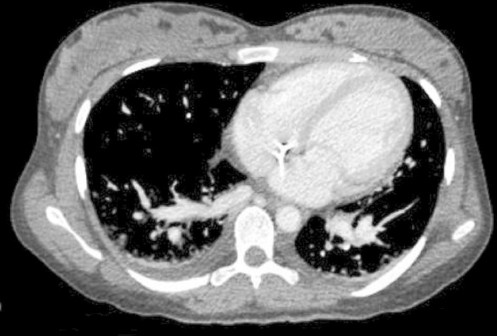
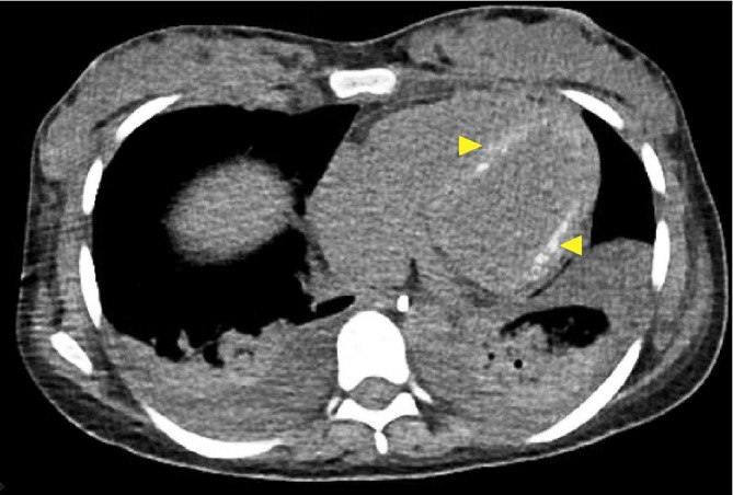
Myocardial calcification is a rare and life-threatening condition. It has been associated with a myriad of causes, including ischaemic heart disease, cardiac surgery, rheumatic fever, and myocarditis. However, this entity is less well recognised in the setting of toxic shock syndrome. Published medical literature is scarce with regard to the pathogenesis and clinical implications of this potential association. We chronicle here the case of a patient with myocardial calcification secondary to toxic shock syndrome from our clinical experience. Furthermore, a systematic literature search of the medical databases PubMed and Google Scholar was conducted. A total of 17 cases fulfilled the inclusion criteria. The data on patients' characteristics, epidemiology, clinical features, comorbid conditions, diagnosis, clinical course and outcome were collected and analysed. The present review outlines our current understanding of the epidemiology of and risk factors for sepsis-related myocardial calcification, the pathophysiology of this condition and currently available approaches to diagnosis.
Keywords: adult intensive care; cardiovascular medicine; healthcare improvement and patient safety; resuscitation.
© BMJ Publishing Group Limited 2019. No commercial re-use. See rights and permissions. Published by BMJ.
Conflict of interest statement
Competing interests: None declared.
Figures

References
Publication types
MeSH terms
LinkOut - more resources
Full Text Sources
Medical