. 2019 Apr 1;151(4):465-477.
doi: 10.1085/jgp.201812215. Epub 2019 Jan 11.

Efficiency measures the conversion of agonist binding energy into receptor conformational change

Affiliations

Efficiency measures the conversion of agonist binding energy into receptor conformational change

Tapan K Nayak et al. J Gen Physiol. .

Erratum in

Abstract

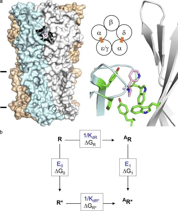
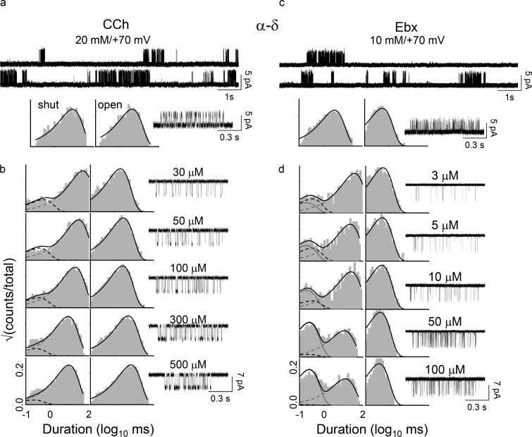
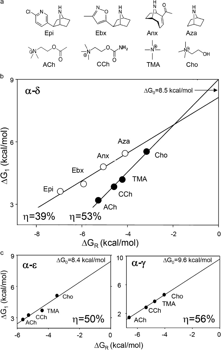
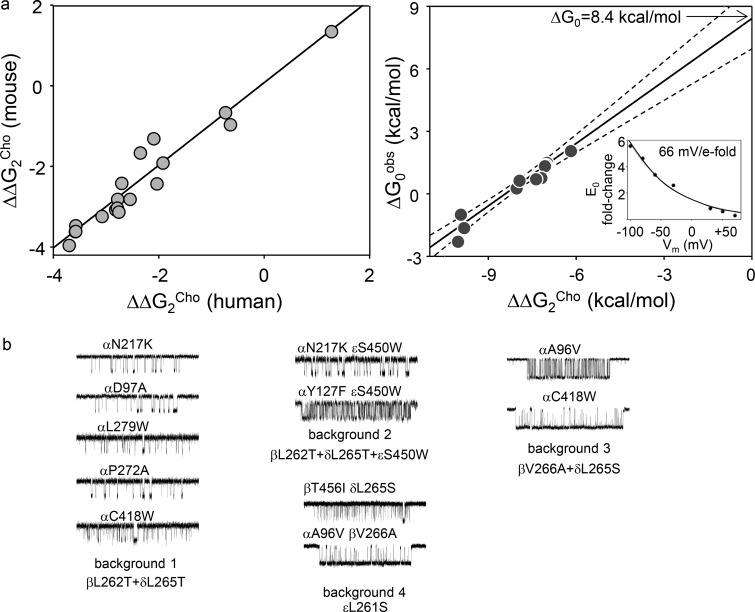
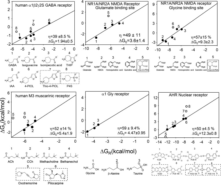
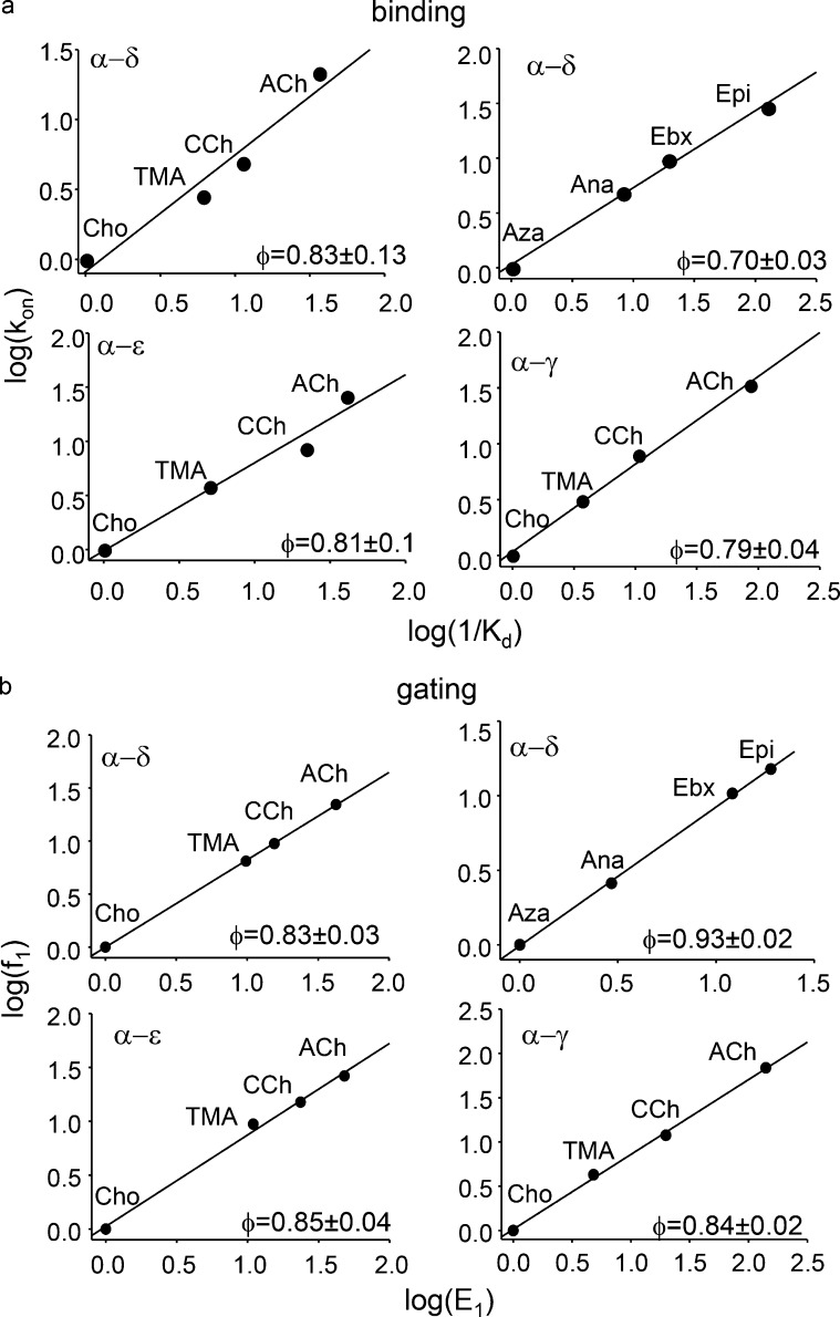
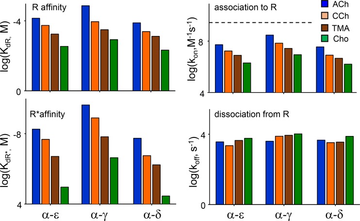
Receptors alternate between resting↔active conformations that bind agonists with low↔high affinity. Here, we define a new agonist attribute, energy efficiency (η), as the fraction of ligand-binding energy converted into the mechanical work of the activation conformational change. η depends only on the resting/active agonist-binding energy ratio. In a plot of activation energy versus binding energy (an "efficiency" plot), the slope gives η and the y intercept gives the receptor's intrinsic activation energy (without agonists; ΔG0). We used single-channel electrophysiology to estimate η for eight different agonists and ΔG0 in human endplate acetylcholine receptors (AChRs). From published equilibrium constants, we also estimated η for agonists of KCa1.1 (BK channels) and muscarinic, γ-aminobutyric acid, glutamate, glycine, and aryl-hydrocarbon receptors, and ΔG0 for all of these except KCa1.1. Regarding AChRs, η is 48-56% for agonists related structurally to acetylcholine but is only ∼39% for agonists related to epibatidine; ΔG0 is 8.4 kcal/mol in adult and 9.6 kcal/mol in fetal receptors. Efficiency plots for all of the above receptors are approximately linear, with η values between 12% and 57% and ΔG0 values between 2 and 12 kcal/mol. Efficiency appears to be a general attribute of agonist action at receptor binding sites that is useful for understanding binding mechanisms, categorizing agonists, and estimating concentration-response relationships.


Figures

Figure 1.
Figure 1.
AChR structure and function. (a) Neurotransmitter binding sites. Left, each site is at a subunit interface (PDB ID 5KXI; Morales-Perez et al., 2016). α-subunit (blue), nicotine (pink), and lines mark approximately the membrane. Middle, each endplate AChR has two neurotransmitter binding sites (ε is adult and γ is fetal). Right, at each site a cluster of aromatic amino acids surrounds the agonist. (b) A cyclic scheme describes receptor operation. Horizontal, agonist binding; vertical, receptor gating. R, resting state (low affinity and closed channel); R*, active state (high affinity and open channel); A, agonist. ΔGR and ΔGR*, binding free energy changes (in direction of arrow) to R and R*; ΔG0 and ΔG1, gating free energy changes with zero and one bound agonist. Corresponding equilibrium constants, blue. Agonists are ligands that bind more strongly to R*.
Figure 2.
Figure 2.
Energy measurements from electrophysiology. The α−δ site of the adult-type human AChRs was studied in isolation after disabling the α−ε site by adding the mutation εP121R. (a) Gating with CCh. Top: Gating with CCh. [CCh] = 20 mM (to fully saturate the α−δ site) and Vm = +70 mV (to reduce channel block by CCh). Openings (top) are clustered; intercluster gaps reflect desensitization and intracluster intervals mainly reflect AR⇄AR* gating. Intracluster interval duration histograms (bottom) and an example cluster. (b) CCh binding. Association and dissociation rate constants were estimated by fitting across [CCh] (see Materials and methods). (c and d) Ebx gating and binding. Free energies were calculated from the equilibrium constants estimated from the forward/backward rate constant ratios.
Figure 3.
Figure 3.
Efficiency plots for human AChR-binding sites. (a) Agonists. Epi, epibatidine; Ebx, epiboxidine; Anx, anatoxin; Aza, azabicycloheptane; ACh, acetylcholine; CCh, carbamylcholine; TMA, tetrmethylammonium; Cho, choline. (b) Efficiency plot for the AChR α−δ neurotransmitter binding site. The y-axis is the gating free energy change and the x-axis is the binding free energy change. The line is the fit by Eq. 3, with energy efficiency (η) calculated from the slope and intrinsic gating energy (ΔG0) from the y intercept. ACh-class agonists are more efficient than Epi-class agonists. (c) Efficiency plots for α−ε and α−γ sites. ACh-class agonists are most efficient at α−γ. The intrinsic gating energy of adult-type AChRs (with an ε subunit) is less positive (more favorable) than of fetal-type (with a γ subunit) AChRs.
Figure 4.
Figure 4.
Intrinsic gating of human AChRs. (a) Left: Mutations far from the binding sites produce similar changes in the diliganded gating energy with Cho (ΔΔG2Cho) in mouse and human AChRs (slope, 1.0 ± 0.1; R2 = 0.95). Each symbol is a different mutation. Right: In adult-type human AChRs, ΔΔG2Cho is caused exclusively by a change in the unliganded gating energy (ΔG0obs; slope = 1.0 ± 0.1, R2 = 0.91; dashed lines, 95% confidence limits). The y intercept (no change in ΔΔG2Cho) is ΔG0 in the WT. Inset: Voltage dependence of E0 in adult-type human AChRs. (b) Example unliganded single-channel current clusters from mutations added to four different background constructs. The clusters (top to bottom) and the backgrounds (left to right) are arranged with increasing open-channel probability (excluding long openings).
Figure 5.
Figure 5.
Efficiency plots for other receptors. In each panel, top is the efficiency plot and bottom is the agonist structures. Energies were calculated from literature values (see text for citations). Gray symbols and boxed ligands are agonists having a different efficiency from the main group, identified statistically and excluded from the linear fit. ΔG0 is kilocalories per mole.
Figure 6.
Figure 6.
Human AChRs REFERs. The slope (φ) of each REFER reports the extent to which a change in equilibrium constant is caused by a change in the forward versus backward rate constant. (a) Binding to the resting state. kon (M−1s−1), association rate constant; KdR, equilibrium dissociation constant. At all sites, agonists differ mainly with regard to association rate constant (ACh-class more so than Epi-class agonists). (b) Gating with one bound agonist. f1 (s−1), forward, channel-opening rate constant; E1, monoliganded gating equilibrium constant. At all sites, agonists differ mainly with regard to the channel-opening rate constant (Epi-class more so than ACh-class agonists).
Figure 7.
Figure 7.
Summary of human AChR-binding constants. Left: Equilibrium constants. For all ACh-class agonists, resting- and active-state–binding energies are greater at the fetal α−γ site than at the adult α−ε and α−δ sites. Right: Rate constants. For all agonists, association to R is slower than diffusion (dashed line, 5 × 10⁹ M−1s−1) and greatest at α−γ. Dissociation rate constants are similar for all agonists and at all sites.

References

    1. Atkinson A. 1994. Fast very robust methods for the detection of multiple outliers. J. Am. Stat. Assoc. 89:1329–1339. 10.1080/01621459.1994.10476872 - DOI
    1. Auerbach A. 1991. Single-channel dose-response studies in single, cell-attached patches. Biophys. J. 60:660–670. 10.1016/S0006-3495(91)82095-1 - DOI - PMC - PubMed
    1. Auerbach A. 2013. The energy and work of a ligand-gated ion channel. J. Mol. Biol. 425:1461–1475. 10.1016/j.jmb.2013.01.027 - DOI - PMC - PubMed
    1. Auerbach A. 2016. Dose–response analysis when there is a correlation between affinity and efficacy. Mol. Pharmacol. 89:297–302. 10.1124/mol.115.102509 - DOI - PMC - PubMed
    1. Brejc K., van Dijk W.J., Klaassen R.V., Schuurmans M., van Der Oost J., Smit A.B., and Sixma T.K.. 2001. Crystal structure of an ACh-binding protein reveals the ligand-binding domain of nicotinic receptors. Nature. 411:269–276. 10.1038/35077011 - DOI - PubMed

Publication types

Associated data