Review
. 2019 Feb 22;294(8):2949-2960.
doi: 10.1074/jbc.REV118.005604. Epub 2019 Jan 10.

Allosteric disulfides: Sophisticated molecular structures enabling flexible protein regulation

Affiliations
Review

Allosteric disulfides: Sophisticated molecular structures enabling flexible protein regulation

Joyce Chiu et al. J Biol Chem. .

Abstract

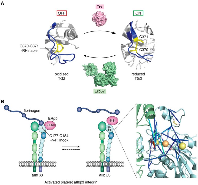
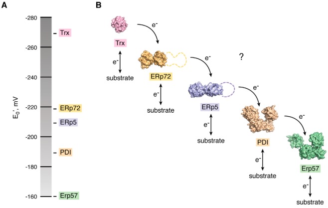
Protein disulfide bonds link pairs of cysteine residues in polypeptide chains. Many of these bonds serve a purely structural or energetic role, but a growing subset of cleavable disulfide bonds has been shown to control the function of the mature protein in which they reside. These allosteric disulfides and the factors that cleave these bonds are being identified across biological systems and life forms and have been shown to control hemostasis, the immune response, and viral infection in mammals. The discovery of these functional disulfides and a rationale for their facile nature has been aided by the emergence of a conformational signature for allosteric bonds. This post-translational modification mostly occurs extracellularly, making these chemical events prime drug targets. Indeed, a membrane-impermeable inhibitor of one of the cleaving factors is currently being trialed as an antithrombotic agent in cancer patients. Allosteric disulfides are firmly established as a sophisticated means by which a protein's shape and function can be altered; however, the full scope of this biological regulation will not be realized without new tools and techniques to study this regulation and innovative ways of targeting it.

Keywords: disulfide; protein chemistry; protein conformation; protein dynamic; protein disulfide isomerase; drug discovery; oxidation-reduction (redox); post-translational modification; protein chemical modification; thiol; allosteric; cysteine; cystine; disulfide bond; oxidoreductase.


Conflict of interest statement

The authors declare that they have no conflicts of interest with the contents of this article

Figures

Figure 1.
Figure 1.
Disulfide bonds form as proteins fold. Protein folding is associated with reduction in conformational entropy that is overcome by favorable enthalpic contributions and by an increase in solvent entropy. Folding of a model protein (blue ribbon) containing disulfide bonds (yellow spheres) is shown. The intermediate states contain zero, one, or two disulfide bonds. The native functional protein contains three disulfide bonds.
Figure 2.
Figure 2.
Mechanisms of cleavage of allosteric disulfides. Disulfide cleavage is a highly directional chemistry. The sulfur ion nucleophile of the oxidoreductase and the two sulfur atoms of the substrate disulfide bond must be in-line for cleavage to proceed. A, cleavage by an oxidoreductase. An active-site sulfur ion of the oxidoreductase attacks one of the allosteric bond sulfur atoms. An intermediate bond between the attacked and the attacking sulfur atoms decomposes and leaves the allosteric disulfide bond reduced and the oxidoreductase active-site sulfurs oxidized. B, cleavage by thiol/disulfide exchange. A conformational change brings an unpaired sulfur ion nucleophile in-line with an allosteric disulfide. The sulfur ion attacks the disulfide bond, cleaving it. C, cleavage by hydrolysis. This mechanism results in formation of a cysteine sulfenic acid and thiol. This mechanism has been observed in one instance, but the biological significance remains to be determined.
Figure 3.
Figure 3.
Allosteric disulfides in transglutaminase-2 and αIIbβ3 integrin. A, Cys-370–Cys-371 disulfide is a switch that turns TG2 “on” (reduced) or “off” (oxidized) in the extracellular matrix. The bond is reduced by thioredoxin and oxidized by ERp57. Control of enzyme activity is via conformational transitions in the protein upon cleavage or formation of the disulfide. The disulfide cysteines are in yellow, and local conformational changes are highlighted in blue ribbon. The oxidized TG2 structure is of PDB identifier 2q3z (42) and reduced structure of 1kv3 (105). The surface representation of the thioredoxin and ERp57 structures are of PDB identifiers 1aiu (106) and 3f8u (107), respectively. B, ERp5 (red oval) triggers fibrinogen release from activated platelet αIIbβ3 integrin by cleaving the βI-domain (cyan oval) Cys-177–Cys-184 disulfide bond. Molecular dynamics simulations show that cleavage of the disulfide results in long-range allosteric effects within the βI-domain, including in the metal-binding sites that are critical for fibrinogen binding. The blue sticks represent the allosteric signaling network measured by force distribution analysis (51). The network involves both cysteines, Asp-119 that is critical for ligand binding, and Asp-217 and Asn-214 that are involved in positioning of the calcium (yellow spheres) and magnesium (orange sphere) ions. The αIIb β-propeller domain is the green cartoon and the βI-domain the cyan cartoon.
Figure 4.
Figure 4.
Possible redox chain of the vascular thiol isomerases. The vascular thiol isomerases that include PDI, ERp57, ERp5, and ERp72 are required for normal thrombosis in mice. A, standard redox potentials of the vascular thiol isomerases (Table 2). B, it is possible that the vascular thiol isomerases function as a redox chain delivering electrons to each other and the substrate disulfides. Their relative redox potentials are consistent with this notion. The surface representations of the oxidoreductases are from the following PDB identifiers: 1aiu for thioredoxin (106); 3idv for ERp72 (108); 3w8j (a-domain (109)) and 4gwr (a′-domain) for ERp5; 4ekz for PDI (102); and 3f8u for ERp57 (107). The dashed lines in ERp72 and ERp5 represent an unknown structure.
Figure 5.
Figure 5.
Classification of disulfide bonds and allosteric conformations. A, classification of disulfide bonds using the five dihedral angles of the cystine residue (center). There are 20 possible disulfide conformations using these angles, and example structures of each from the PDB are shown. B, incidence of the three allosteric disulfide conformations. The −RHstaple, −LHhook, and −/+RHhook conformations constitute ∼20% of all nonredundant disulfide bonds in the PDB, but ∼80% of the structurally defined allosteric bonds (n = 32 (61)). C, allosteric −RHstaple and −/+RHhook conformations are naturally strained. The pairwise forces between the cysteine residues of the 20 different disulfide conformations were calculated using force distribution analysis (65). The stresses on the −RHstaple and −/+RHhook disulfide bonds are significantly higher than the other 18 conformations. The bonded pre-stress (or tensile pre-stress) measure comprises all force terms involving the sulfur atoms of the disulfide, so is the relevant indicator for disulfide reactivity. Mean tensile pre-stress of the 20 disulfide conformations was adapted from Fig. 2D of Zhou et al. (65). D, tensile pre-stress of the −RHstaple and −/+RHhook bonds is a result of stretching of the sulfur–sulfur bond length (indicated by d) and neighboring α angles.

Similar articles

Cited by

References

    1. Wong J. W., Ho S. Y., and Hogg P. J. (2011) Disulfide bond acquisition through eukaryotic protein evolution. Mol. Biol. Evol. 28, 327–334 10.1093/molbev/msq194 - DOI - PubMed
    1. Brooks D. J., Fresco J. R., Lesk A. M., and Singh M. (2002) Evolution of amino acid frequencies in proteins over deep time: inferred order of introduction of amino acids into the genetic code. Mol. Biol Evol. 19, 1645–1655 10.1093/oxfordjournals.molbev.a003988 - DOI - PubMed
    1. Jordan I. K., Kondrashov F. A., Adzhubei I. A., Wolf Y. I., Koonin E. V., Kondrashov A. S., and Sunyaev S. (2005) A universal trend of amino acid gain and loss in protein evolution. Nature 433, 633–638 10.1038/nature03306 - DOI - PubMed
    1. Schmidt B., Ho L., and Hogg P. J. (2006) Allosteric disulfide bonds. Biochemistry 45, 7429–7433 10.1021/bi0603064 - DOI - PubMed
    1. Hogg P. J. (2003) Disulfide bonds as switches for protein function. Trends Biochem. Sci. 28, 210–214 10.1016/S0968-0004(03)00057-4 - DOI - PubMed

Publication types

Substances