. 2019 Jan 29;116(5):1686-1691.
doi: 10.1073/pnas.1812155116. Epub 2019 Jan 11.

PET imaging of microglia by targeting macrophage colony-stimulating factor 1 receptor (CSF1R)

Affiliations

PET imaging of microglia by targeting macrophage colony-stimulating factor 1 receptor (CSF1R)

Andrew G Horti et al. Proc Natl Acad Sci U S A. .

Abstract

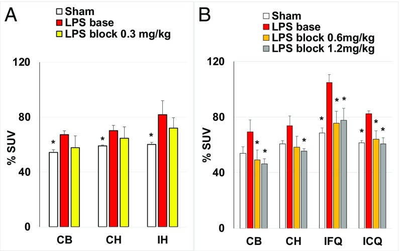
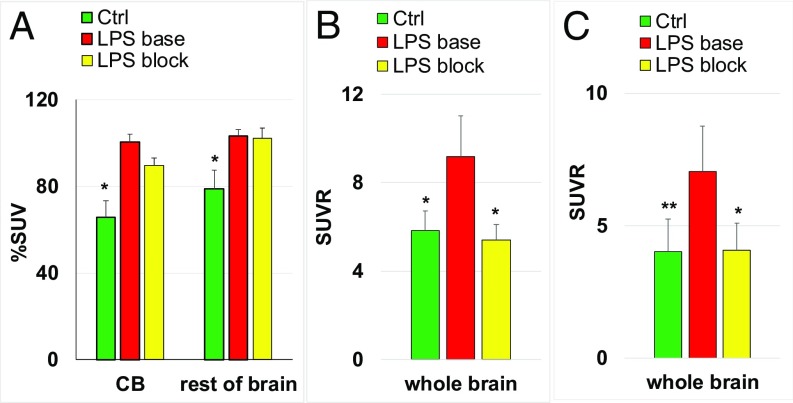
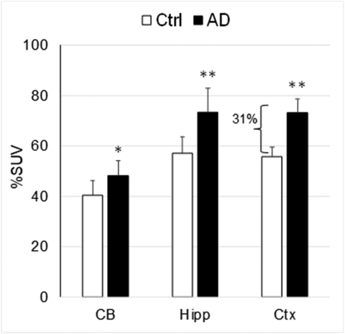
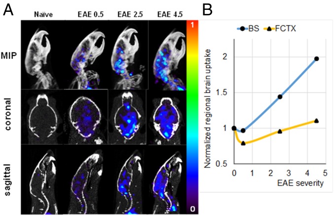
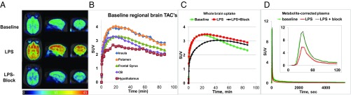
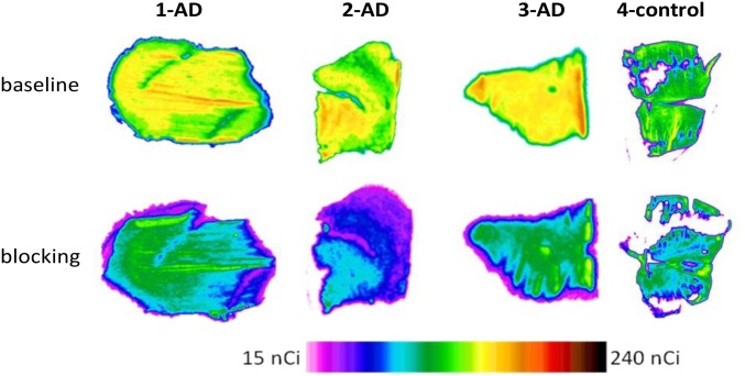
While neuroinflammation is an evolving concept and the cells involved and their functions are being defined, microglia are understood to be a key cellular mediator of brain injury and repair. The ability to measure microglial activity specifically and noninvasively would be a boon to the study of neuroinflammation, which is involved in a wide variety of neuropsychiatric disorders including traumatic brain injury, demyelinating disease, Alzheimer's disease (AD), and Parkinson's disease, among others. We have developed [11C]CPPC [5-cyano-N-(4-(4-[11C]methylpiperazin-1-yl)-2-(piperidin-1-yl)phenyl)furan-2-carboxamide], a positron-emitting, high-affinity ligand that is specific for the macrophage colony-stimulating factor 1 receptor (CSF1R), the expression of which is essentially restricted to microglia within brain. [11C]CPPC demonstrates high and specific brain uptake in a murine and nonhuman primate lipopolysaccharide model of neuroinflammation. It also shows specific and elevated uptake in a murine model of AD, experimental allergic encephalomyelitis murine model of demyelination and in postmortem brain tissue of patients with AD. Radiation dosimetry in mice indicated [11C]CPPC to be safe for future human studies. [11C]CPPC can be synthesized in sufficient radiochemical yield, purity, and specific radioactivity and possesses binding specificity in relevant models that indicate potential for human PET imaging of CSF1R and the microglial component of neuroinflammation.

Keywords: CSF1R; DAM; [11C]CPPC; neuroinflammation; positron-emission tomography.


Conflict of interest statement

The authors declare no conflict of interest.

Figures

Fig. 1.
Fig. 1.
Comparison of [11C]CPPC brain uptake in sham and LPS: right forebrain injected mice, baseline, and blocking. Two independent experiments (A and B) were performed. The time point was 45 min after radiotracer injection; LPS (5 µg in 0.5 µL) or saline (0.5 µL) was injected into the right forebrain (ipsilateral frontal quadrant) 2–3 d before the radiotracer study. Blocker (CPPC) was injected i.p. 5 min before the radiotracer. (A) The regions of interest (ROIs) are cerebellum (CB), ipsilateral hemisphere (IH), and contralateral hemisphere (CH). The data are mean %SUV ± SD (n = 3). (B) The ROIs are cerebellum (CB), contralateral hemisphere (CH), ipsilateral caudal quadrant (ICQ), and ipsilateral frontal quadrant (IFQ). The data are mean %SUV ± SD (n = 4). Statistical analysis: comparison of LPS-baseline versus sham or LPS-block. *P < 0.05; no asterisk indicates P > 0.05 (ANOVA).
Fig. 2.
Fig. 2.
Brain uptake of CSF1R radiotracer [11C]CPPC in control (Ctrl), LPS (i.p.)-treated mice (LPS base), and LPS (i.p.)-treated mice plus blocking with CSF1R inhibitors (LPS block) in three independent experiments. The time point was 45 min after radiotracer injection [LPS (10 mg/kg)]. (A) Data are mean %SUV ± SD (n = 5). CB, cerebellum. (B) Data are mean SUVR ± SD (n = 5). Blocker (CPPC, 1 mg/kg, i.p.) was injected in the LPS-treated mice. (C) Data are mean SUVR ± SD (n = 3–6). Blocker (compound 8, 2 mg/kg, i.p.) was injected in the LPS-treated mice. Statistical analysis: comparison of LPS-baseline versus control or LPS-block. *P < 0.01; **P = 0.03; no asterisk indicates P > 0.05 (ANOVA).
Fig. 3.
Fig. 3.
Comparison of the [11C]CPPC brain uptake in transgenic AD (n = 6) and control (n = 5) mice. Time-point – 45 min after radiotracer injection. Data: mean %SUV ± SD. *P = 0.04, **P < 0.005 (ANOVA). The uptake of [11C]CPPC was significantly greater in AD mouse brain regions. CB, cerebellum; Ctx, cortex; Hipp, hippocampus.
Fig. 4.
Fig. 4.
[11C]CPPC PET/CT imaging in murine EAE. (A) MIP (Top), coronal (Middle), and sagittal (Bottom) slices showing radiotracer uptake from 45 to 60 min per projection in the indicated mice. Color scale range shows %ID/g tissue. (B) Regional brain uptake normalized by uptake in control animal vs. EAE severity. BS, brainstem; FCTX, frontal cortex.
Fig. 5.
Fig. 5.
PET imaging of [11C]CPPC in the same baboon in baseline, LPS, and LPS-plus-blocking experiments. The LPS dose was 0.05 mg/kg (i.v.), 4 h before radiotracer injection. (A) Parametric (VT) images. (B) Baseline regional brain SUV time-uptake curves of [11C]CPPC. (C) Whole-brain SUV time-uptake curves of [11C]CPPC: baseline (green), after LPS treatment (red) and blocking after LPS treatment (black). (D) Metabolite-corrected plasma SUV time-uptake curves of [11C]CPPC: baseline (green), after LPS treatment (red), and LPS-plus-blocking (black). The Inset in D shows first 120 s of scanning.
Fig. 6.
Fig. 6.
Postmortem human autoradiography/[11C]CPPC images (baseline and blocking) in inferior parietal lobe gray matter slices. Three subjects with Alzheimer’s disease (1-AD, 2-AD, and 3-AD) and control (4-control) subject. See also SI Appendix, Fig. S14 and Tables S5 and S6.

References

    1. Masgrau R, Guaza C, Ransohoff RM, Galea E. Should we stop saying ‘glia’ and ‘neuroinflammation’? Trends Mol Med. 2017;23:486–500. - PubMed
    1. Tronel C, et al. Molecular targets for PET imaging of activated microglia: The current situation and future expectations. Int J Mol Sci. 2017;18:E802. - PMC - PubMed
    1. Janssen B, Vugts DJ, Windhorst AD, Mach RH. PET imaging of microglial activation-beyond targeting TSPO. Molecules. 2018;23:607. - PMC - PubMed
    1. Aguzzi A, Barres BA, Bennett ML. Microglia: Scapegoat, saboteur, or something else? Science. 2013;339:156–161. - PMC - PubMed
    1. Akiyama H, et al. Expression of the receptor for macrophage colony stimulating factor by brain microglia and its upregulation in brains of patients with Alzheimer’s disease and amyotrophic lateral sclerosis. Brain Res. 1994;639:171–174. - PubMed

Publication types

Substances