. 2019 Mar 7;9(3):737-748.
doi: 10.1534/g3.118.200975.

Conditional Synaptic Vesicle Markers for Drosophila

Affiliations

Conditional Synaptic Vesicle Markers for Drosophila

Jessica L Williams et al. G3 (Bethesda). .

Abstract

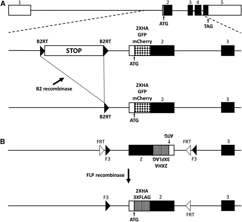
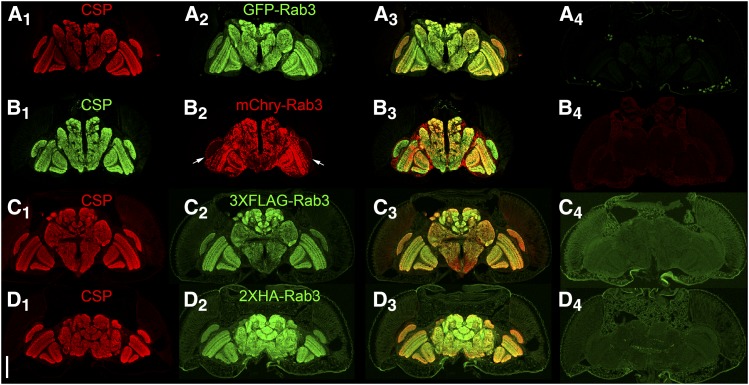
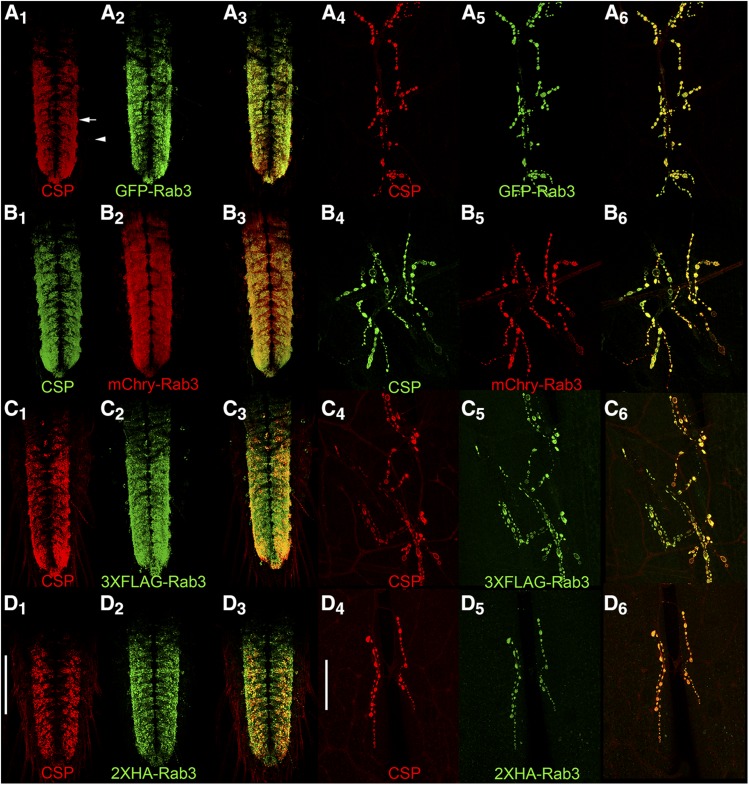
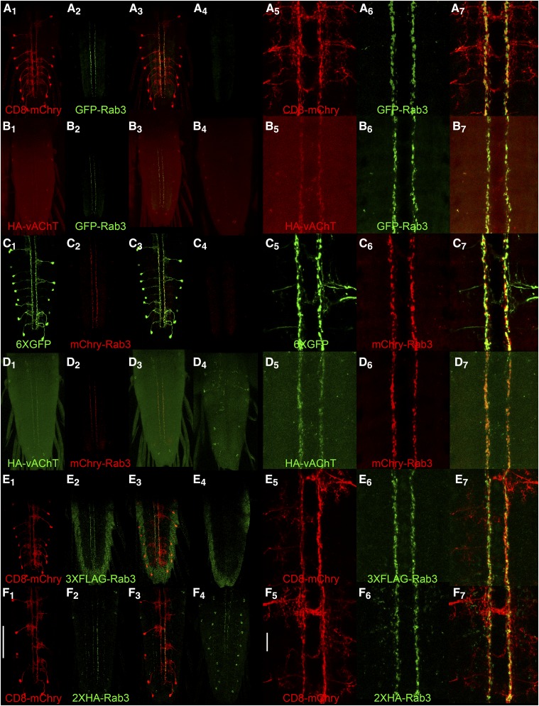
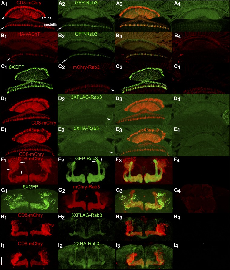
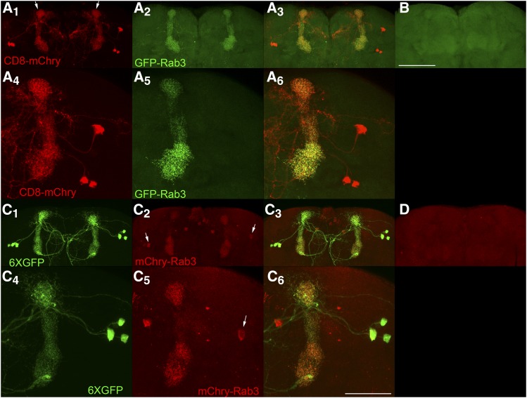
The release of neurotransmitters from synaptic vesicles (SVs) at pre-synaptic release sites is the principle means by which information transfer between neurons occurs. Knowledge of the location of SVs within a neuron can thus provide valuable clues about the location of neurotransmitter release within a neuron and the downstream neurons to which a given neuron is connected, important information for understanding how neural circuits generate behavior. Here the development and characterization of four conditional tagged SV markers for Drosophila melanogaster is presented. This characterization includes evaluation of conditionality, specificity for SV localization, and sensitivity of detection in diverse neuron subtypes. These four SV markers are genome-edited variants of the synaptic vesicle-specific protein Rab3. They depend on either the B2 or FLP recombinases for conditionality, and incorporate GFP or mCherry fluorescent proteins, or FLAG or HA epitope tags, for detection.

Keywords: Drosophila; conditional; epitope; fluorescent; synaptic vesicle.


Figures

Figure 1
Figure 1
Strategies for generating conditional tagged variants of a Drosophila Rab3 synaptic vesicle marker. A) top: genomic exon structure of Drosophila Rab3. middle: strategy 1 involves incorporating a B2 recombinase target (B2RT)-flanked transcription STOP cassette upstream of exon 2 and either a 2XHA epitope tag, or a GFP or mCherry fluorescent protein tag, at the amino-terminus of Rab3. Prior to excision of the transcription STOP cassette, the tagged variants of Rab3 will not be expressed. bottom: after B2 recombinase expression in neurons of interest, the tagged variants of Rab3 will be expressed in neurons in which the STOP cassette has been excised. B) top: strategy 2 involves paired FRT and F3 FLP recombinase target sites flanking an inverted exon 2 containing 2XHA or 3XFLAG amino-terminal epitope tags. bottom: after FLP recombinase expression in neurons of interest and an odd number of inversions followed by an excision, the tagged variants of Rab3 will be stably expressed. Note that FLP recombinase is functional with FRT pairs and F3 pairs but is not functional in an FRT-F3 combination.
Figure 2
Figure 2
Assessment of co-localization between the endogenous synaptic vesicle marker Cysteine String Protein (CSP) and the tagged variants of Rab3 in immunostained cryostat sections of the adult brain after germline excision (A, B), or germline inversion (C, D). A1-D1) CSP localizes to the neuropil; A2) GFP-Rab3 shows a neuropil distribution; B2) mCherry-Rab3 shows a neuropil distribution except for very weak expression in the lamina (arrows); C2) 3XFLAG-Rab3 and D2) 2XHA-Rab3 show a neuropil distribution; A3-D3) Overlay. A4-D4) Controls without a GAL4 driver exhibit minimal levels of background fluorescence. A4-D4 were processed and imaged identically to A2-D2. Scale bar: 100μm.
Figure 3
Figure 3
Assessment of co-localization via immunostaining between the endogenous synaptic vesicle marker Cysteine String Protein (CSP) and the tagged variants of Rab3 in third instar larva after pan-neuronal recombinase expression. In the ventral nerve cord (A1-3-D1-3) and at the neuromuscular junction (A4-6-D4-6), CSP exhibits a high degree of co-localization with GFP-Rab3 (A), mCherry-Rab3 (B), 3XFLAG-Rab3 (C), and 2XHA-Rab3 (D). Scale bars: 100μm (D1); 50μm (D4).
Figure 4
Figure 4
Conditional expression of tagged Rab3 variants in Basin-4 larval neurons. A1, C1, E1, F1) The location and structure of Basin-4 neurons in the larval ventral nerve cord visualized with the plasma membrane marker CD8-mcherry or the cytoplasmic marker 6XGFP. There are two symmetric Basin-4 neurons per segment with peripherally located cell bodies and branched axodendritic processes extending medially to the central region of the VNC. A2, C2, E2, F2) Each of the tagged Rab3 variants exhibits highly restricted localization to two longitudinal strips near the center of the VNC when conditionally expressed in Basin-4 neurons. A3, C3, E3, F3) Overlay. A4, C4) Background levels of expression of GFP-Rab3 and mCherry-Rab3 in the absence of a GAL4 driver. A4 and C4 were imaged and processed identically to A2 and C2, respectively. E4, F4) Background levels of signal detected with anti-FLAG and anti-HA antibodies in negative controls. E4 and F4 were imaged and processed identically to E2 and F2, respectively. A5, C5, E5, F5) High magnification images of the central region of the VNC of Basin-4 neurons visualized with CD8-mCherry or 6XGFP. A6, C6, E6, F6) High magnification images of the tagged Rab3 variants in the central region of the VNC of Basin-4 neurons. A7, C7, E7, F7) Overlay. The tagged Rab3 variants are excluded from all regions of Basin-4 neurons except for two central longitudinal strips. B1, D1) The cholinergic synaptic vesicle marker HA-vAChT localizes to two central strips in the VNC upon conditional expression in Basin-4 neurons. B2, D2) GFP-Rab3 and mCherry-Rab3 localization is restricted to two central strips in the VNC upon conditional expression in Basin-4 neurons. B3, D3) Overlay. HA-vAChT co-localizes with GFP-Rab3 and mCherry-Rab3 in two central strips of Basin-4 neurons. B4, D4) Background levels of expression of HA-vAChT in the absence of a GAL4 driver. B4 and D4 were imaged and processed identically to B2 and D2, respectively. B5, D5) High magnification images of of HA-vAChT conditional expression in Basin-4 neurons in the central region of the VNC. B6, D6) High magnification images of of GFP-Rab3 and mCherry-Rab3 conditional expression in Basin-4 neurons in the central region of the VNC. B7, D7) Overlay. HA-vAChT exhibits nearly indistinguishable localization with GFP-Rab3 and mCherry-Rab3 in Basin-4 neurons at high magnification. Scale bars: 100μm (F1); 15μm (F5).
Figure 5
Figure 5
Conditional expression of tagged Rab3 variants in adult L2 lamina optic lobe neurons and MB131B adult γ-lobe mushroom body neurons. A1, C1, D1, E1) Anatomy of L2 lamina neurons visualized with the plasma membrane marker CD8-mCherry or the cytoplasmic marker 6XGFP. The structure of L2 lamina neurons includes cell bodies located between photoreceptor rhabdomeres and the lamina neuropil (arrowhead, A1), axodendritic processes that extend through the lamina neuropil, and synaptic terminals in the medulla (arrow, A1). A2, C2, D2, E2) Localization of tagged Rab3 variants in L2 lamina neurons. Arrows indicate synaptic terminals in the medulla. A3, C3, D3, E3) Overlay. A4, C4, D4, E4) Controls for the tagged Rab3 variants in the absence of a GAL4 driver showing background levels of autofluorescence due to w+ pigment present in photoreceptors. The significant levels of autofluorescence in the lamina, especially in the green channel, confound assessment of localization in the lamina neuropil. Signal above background is detectable in L2 synaptic terminals for all four tagged Rab3 variants, although it is noticeably weaker for 3XFLAG-Rab3 and HA-Rab3. B1-3) Conditional co-expression of HA-vAChT (B1) and GFP-Rab3 (B2) in L2 lamina neurons. Arrows indicate synaptic terminals. B3) Overlay. HA-vAChT and GFP-Rab3 show a high degree of co-localization in L2 synaptic terminals. B4) Control for HA-vAChT in the absence of a GAL4 driver. F1, G1, H1, I1). Anatomy of MB131B Kenyon cell neurons γ-lobe mushroom body neurons visualized with CD8-mCherry or 6XGFP. For one lobe in F1, cell bodies are indicated with an arrow, axodendritic processes with a small arrowhead, and γ-lobe neuropil with a large arrowhead. F2, G2, H2, I2) Localization of tagged Rab3 variants in MB131B mushroom body neurons. F3, G3, H3, I3) Overlay. All tagged Rab3 variants localize to the γ-lobe neuropil where synaptic terminals are located, but not to the cell body or axodendritic regions of MB131B neurons with GFP-Rab3 and mCherry-Rab3 showing significantly stronger signal than 3XFLAG-Rab3 or 2XHA-Rab3. In addition, all tagged variants localize to mushroom body α- and α’-lobe neuropil and variably in β-lobe neuropil. In F2 for one lobe the α-lobe is indicated with a large arrowhead, the α’-lobe with an arrow and the β-lobe with a small arrowhead. F4, G4, H4, I4) Controls for the tagged Rab3 variants in the absence of a GAL4 driver imaged and processed identically to their corresponding variants shown in F2-I2. The absence of signal in mushroom body neuropil in F4-I4 suggests the signal in mushroom body neuropil outside the γ-lobe in F2-I2 is due to developmental expression of the MB131B GAL4 driver in neurons that comprise the other neuropils and is not due to constitutive/leaky expression from the tagged Rab3 variants. Scale bars: 50μm (E1 and I1).
Figure 6
Figure 6
Conditional expression of GFP-Rab3 and mCherry-Rab3 in dopaminergic PPL1-α3 neurons. A1, A4, C1, C4) The PPL1-α3 neurons are represented by the MB060B GAL4 driver that expresses in three neurons of each brain hemisphere as visualized with the plasma membrane marker CD8-mCherry or the cytoplasmic marker 6XGFP. PPL1-α3 neurons extend axodendritic processes that project medially from their cell bodies primarily to the α-lobe of the mushroom body (arrows, A1), but also to the midline. A2, A5) Conditional expression of GFP-Rab3 in PPL1-α3 neurons. GFP-Rab3 localizes exclusively to the α-lobe of the mushroom body where synaptic contact sites are located with no discernible expression in cell bodies or processes outside the α-lobe. A5) A higher resolution image reveals the fine scale structure of SV distribution. A3, A6) Overlays. B) Negative control with all genetic components except the GAL4 driver shows background levels of signal. C2, C5) Conditional expression of mCherry-Rab3 in PPL1-α3 neurons. mCherry-Rab3 localizes predominantly to the α-lobe of the mushroom body, although cell body signal in some PPL1-α3 neurons is perceptible (arrows). This likely does not represent actual SVs since no cell body signal was observed with GFP-Rab3. Outside the cell bodies, mCherry-Rab3 localizes exclusively to the α-lobe region of the mushroom body in a distribution almost indistinguishable from GFP-Rab3. C5) A higher resolution image shows the fine scale structure of SV localization. C3, C6) Overlays. D) Negative control with all genetic components except the GAL4 driver shows background levels of signal. These results demonstrate the signal from the GFP-Rab3 and mCherry-Rab3 SV markers is of sufficient strength to reveal the SV distribution in individual neurons. Scale bars 100μm (B); 50μm (C6).

Similar articles

Cited by

References

    1. Aso Y., Hattori D., Yu Y., Johnston R. M., Iyer N. A., et al. , 2014. The neuronal architecture of the mushroom body provides a logic for associative learning. eLife 3: e04577 10.7554/eLife.04577 - DOI - PMC - PubMed
    1. Bassett A. R., Tibbit C., Ponting C. P., Liu J. L., 2013. Highly efficient targeted mutagenesis of Drosophila with the CRISPR/Cas9 system. Cell Reports 4: 220–228. 10.1016/j.celrep.2013.06.020 - DOI - PMC - PubMed
    1. Bushey D., Tononi G., Cirelli C., 2009. The Drosophila fragile X mental retardation gene regulates sleep need. J. Neurosci. 29: 1948–1961. 10.1523/JNEUROSCI.4830-08.2009 - DOI - PMC - PubMed
    1. Certel S. J., Thor S., 2004. Specification of Drosophila motoneuron identity by the combinatorial action of POU and LIM-HD factors. Development 131: 5429–5439. 10.1242/dev.01418 - DOI - PubMed
    1. Dionne H., Hibbard K. L., Cavallaro A., Kao J. C., Rubin G. M., 2018. Genetic Reagents for Making Split-GAL4 Lines in Drosophila. Genetics 209: 31–35. 10.1534/genetics.118.300682 - DOI - PMC - PubMed

Publication types

LinkOut - more resources