. 2019 May;33(5):838-844.
doi: 10.1038/s41433-018-0332-8. Epub 2019 Jan 11.

Non-physician grader reliability in measuring morphological features of the optic nerve head in stereo digital images

Affiliations

Non-physician grader reliability in measuring morphological features of the optic nerve head in stereo digital images

Victoria Addis et al. Eye (Lond). 2019 May.

Abstract

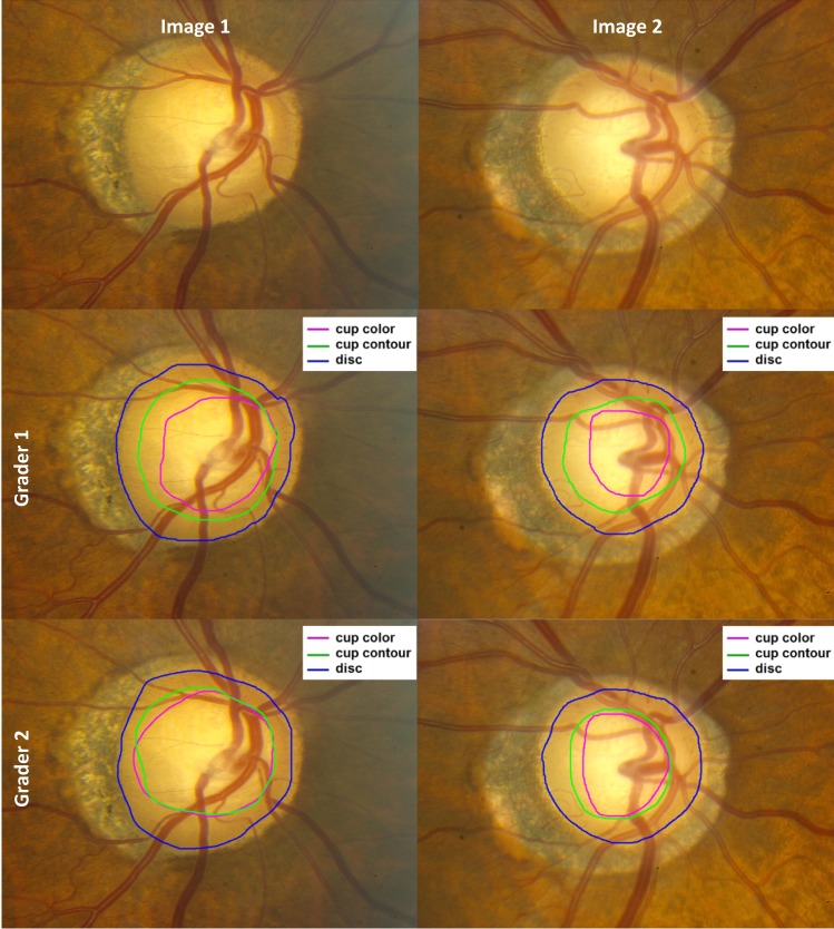
Objective: To introduce a new method of grading optic nerve stereo disc photographs and evaluate reproducibility of assessments by non-physician graders in a reading center.

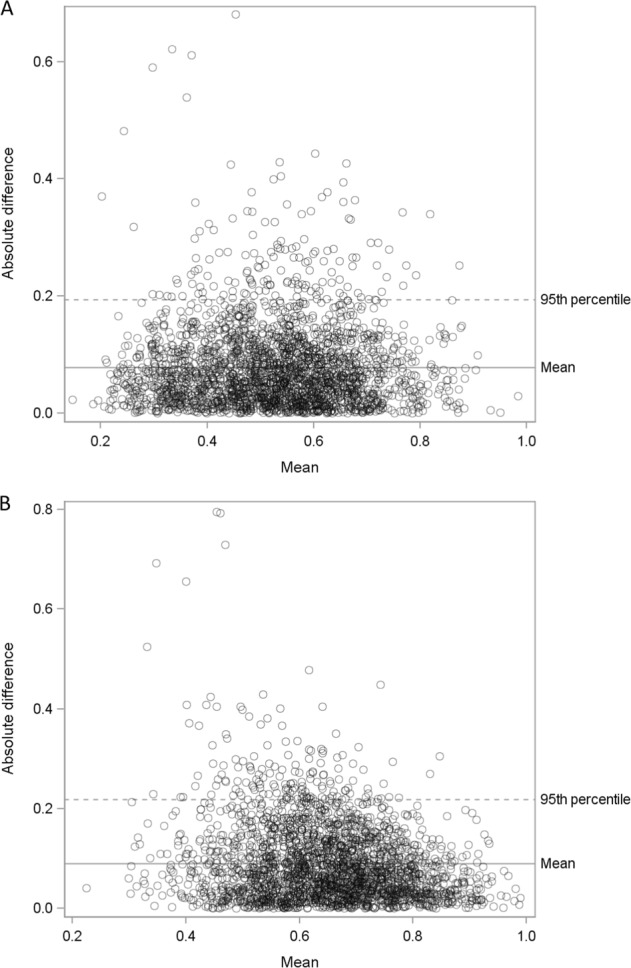
Methods: Three non-physician graders, experienced in grading features of the retina but not the optic nerve head (ONH), were trained by glaucoma specialists to assess digital stereo color images of the ONH. These graders assessed a total of 2554 digital stereo disc images from glaucoma cases and controls participating in the Primary Open-Angle African American Glaucoma Genetics (POAAGG) study by outlining the optic cup and disc. Inter-grader reproducibility of area, height, and width measurements was analyzed.

Results: Among all images, the intraclass correlation (95% confidence interval) was 0.90 (0.89, 0.90) for the cup area using only color cues; 0.92 (0.91, 0.92) for the cup area using contour and vascular cues; and 0.99 (0.99, 0.99) for the optic disc area. The intraclass correlation for cup-to-disc ratio (CDR) was 0.61 (0.58, 0.63), as determined by the ratio of optic cup area to optic disc area (using contour and vascular cues). The CDR difference by graders for area was ≤ 0.1 in 65% of images using color/vascular cues and ≤0.1 in 71% of images using color cues.

Conclusions: After adequate training, non-physician graders were able to measure the optic nerve CDR with high inter-grader reliability.


Conflict of interest statement

The authors declare that they have no conflict of interest.

Figures

Fig. 1
Fig. 1
Examples of stereo disc photos outlined for cup color, cup contour, and disc by two graders. For Image 1, areas were 72,070 (Grader 1) and 94,006 (Grader 2) for cup color; 108,667 (Grader 1) and 98,751 (Grader 2) for cup contour; and 174,421 (Grader 1) and 172,310 (Grader 2) for the disc. CDR was 0.41 (Grader 1) and 0.55 (Grader 2) using cup color, and 0.62 (Grader 1) and 0.57 (Grader 2) using cup contour. For Image 2, areas were 39,859 (Grader 1) and 48,728 (Grader 2) for cup color; 75,195 (Grader 1) and 63,352 (Grader 2) for cup contour; and 135,916 (Grader 1) and 133,663 (Grader 2) for the disc. CDR was 0.29 (Grader 1) and 0.36 (Grader 2) using cup color, and 0.55 (Grader 1) and 0.47 (Grader 2) using cup contour. Area units are unscaled pixels
Fig. 2
Fig. 2
Bland-Altman plots for cup-to-disc ratio for color and contour grading. a Bland-Altman plot of the absolute value of the difference between two graders of the cup-to-disc ratio by the mean of the two values when using the area of the cup as defined by color and the area of the disc. The mean absolute difference was 0.078, and the 95th percentile of the differences is 0.193. b Bland-Altman plot of the absolute value of the difference between two graders of the cup-to-disc ratio by the mean of the two values when using the area of the cup as defined by contour and the area of the disc. The mean absolute difference was 0.089, and the 95th percentile of the differences is 0.218

References

    1. Gandhi M, Dubey S. Evaluation of the optic nerve head in glaucoma. J Curr Glaucoma Pract. 2013;7:106–14. doi: 10.5005/jp-journals-10008-1146. - DOI - PMC - PubMed
    1. Tham YC, Li X, Wong TY, Quigley HA, Aung T, Cheng CY. Global prevalence of glaucoma and projections of glaucoma burden through 2040: a systematic review and meta-analysis. Ophthalmology. 2014;121:2081–90. doi: 10.1016/j.ophtha.2014.05.013. - DOI - PubMed
    1. Spaeth GL, Lopes JF, Junk AK, Grigorian AP, Henderer J. Systems for staging the amount of optic nerve damage in glaucoma: a critical review and new material. Surv Ophthalmol. 2006;51:293–315. doi: 10.1016/j.survophthal.2006.04.008. - DOI - PubMed
    1. Hitchings RA, Spaeth GL. The optic disc in glaucoma. I: Classification. Br J Ophthalmol. 1976;60:778–85. doi: 10.1136/bjo.60.11.778. - DOI - PMC - PubMed
    1. Lichter PR. Variability of expert observers in evaluating the optic disc. Trans Am Ophthalmol Soc. 1976;74:532–72. - PMC - PubMed